4FE3 image
Deposition Date 2012-05-29
Release Date 2012-08-29
Last Version Date 2023-09-13
Entry Detail
PDB ID:
4FE3
Keywords:
Title:
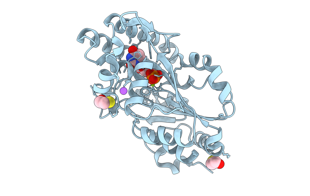
Structure of murine cytosolic 5'-nucleotidase III complexed with uridinine monophosphate
Biological Source:
Source Organism:
Mus musculus (Taxon ID: 10090)
Host Organism:
Method Details:
Experimental Method:
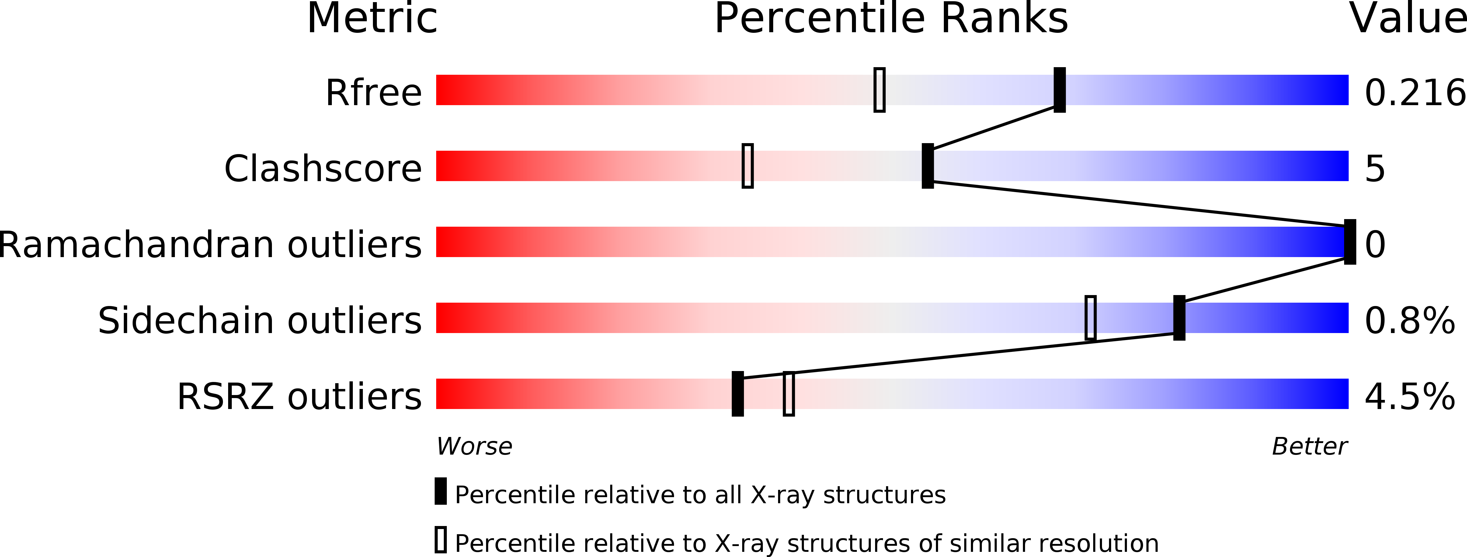
Resolution:
1.74 Å
R-Value Free:
0.21
R-Value Work:
0.16
R-Value Observed:
0.17
Space Group:
P 43 2 2
Macromolecular Entities
Structures with similar UniProt ID
Protein Blast
Polymer Type:polypeptide(L)
Molecule:Cytosolic 5'-nucleotidase 3
Gene (Uniprot):Nt5c3a
Mutagens:D51N
Chain IDs:A
Chain Length:297
Number of Molecules:1
Biological Source:Mus musculus
Primary Citation
Structural Basis of Substrate Specificity and Selectivity of Murine Cytosolic 5'-Nucleotidase III.
J.Mol.Biol. 423 540 554 (2012)
PMID: 22925580 DOI: 10.1016/j.jmb.2012.08.014

Abstact

Cytosolic 5'-nucleotidase III (cN-III) is responsible for selective degradation of pyrimidine 5'-monoribonucleotides during maturation of reticulocytes to erythrocytes. The lack of this enzymatic activity due to genetic aberrations or lead poisoning results in a mild to moderate nonspherocytic hemolytic anemia. In affected individuals, pyrimidine nucleotides as well as their precursor polymers and their off-path metabolites accumulate in erythrocytes, interfering with their proper function in ways that are not yet fully understood. This report describes the first X-ray structure of a catalytically inactivated variant of murine cN-III with a natural substrate, uridine 5'-monophosphate, in the active site at 1.74Å resolution. The structure captures in an atomic detail the closed conformation that cN-III adopts upon substrate binding. Structure and sequence analysis coupled with enzymatic characterization of several mutants confirmed that the aromatic ring of a nitrogenous base of substrate nucleotide is stabilized by parallel π-stacking interactions with conserved aromatic rings of Trp113 and His68. The nitrogenous base is further stabilized by T-shaped stacking with the conserved aromatic ring of Tyr114, as well as by polar contacts with side chains of Thr66 and Ser117. Two water molecules help to stabilize the nucleotide binding by bridging it to protein residues Asp72 and His68 via hydrogen bonds. Finally, fully conserved Glu96 is responsible for recognition of ribose ring via two hydrogen bonds. The presented substrate complex structure elucidates how cN-III achieves specificity for pyrimidine 5'-nucleotides and how it selects against purine 5'-nucleotides.

Legend

Protein

Chemical

Disease

Primary Citation of related structures