4FD9 image
Entry Detail
PDB ID:
4FD9
Title:
Crystal structure of the third beta-gamma-crystallin domain of Crybg3 (betagamma-crystallin domain-containing protein 3) from Mus musculus
Biological Source:
Source Organism:
Host Organism:
PDB Version:
Deposition Date:
2012-05-26
Release Date:
2013-04-10
Method Details:
Experimental Method:
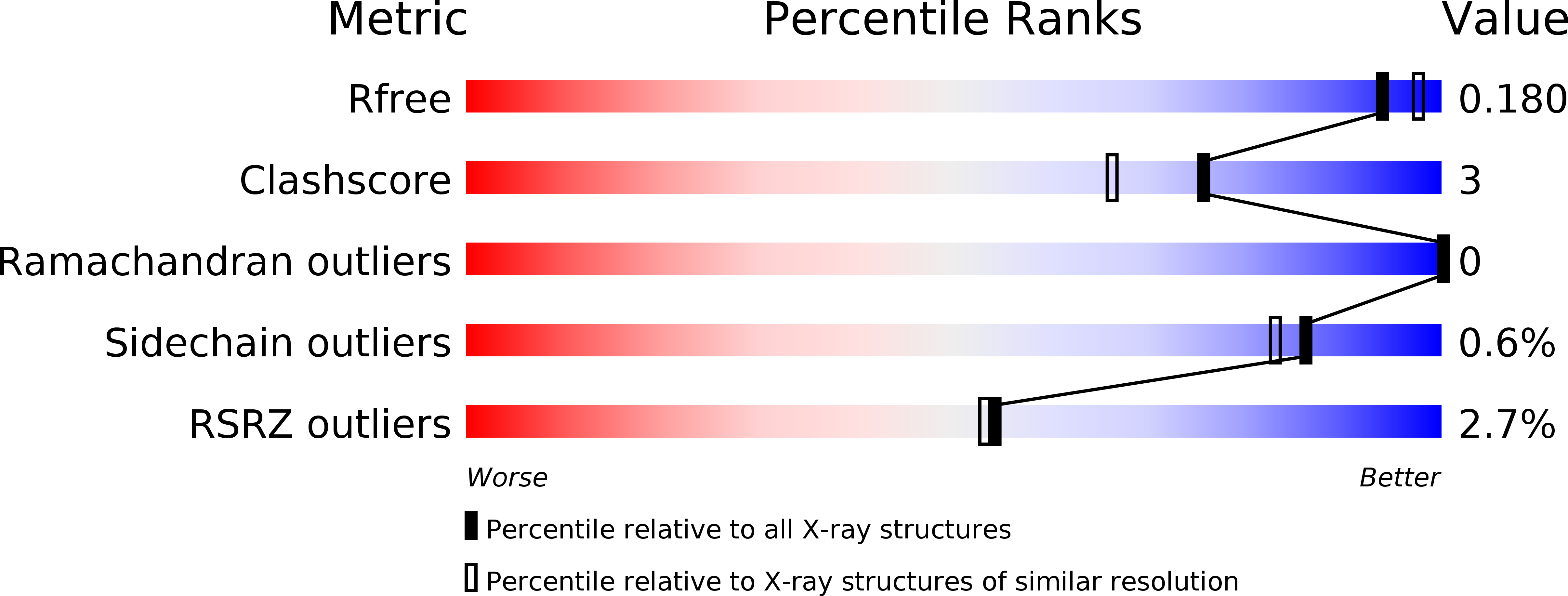
Resolution:
1.86 Å
R-Value Free:
0.17
R-Value Work:
0.13
R-Value Observed:
0.13
Space Group:
P 1 21 1
Macromolecular Entities
Polymer Type:polypeptide(L)
Description:Beta/gamma crystallin domain-containing protein 3
Chain IDs:A, B
Chain Length:92
Number of Molecules:2
Biological Source:Mus musculus
Primary Citation
Aggregation-prone near-native intermediate formation during unfolding of a structurally similar nonlenticular beta/gamma-crystallin domain
Biochemistry 51 8502 8513 (2012)
PMID: 23043265 DOI: 10.1021/bi300844u

Abstact

The folding and unfolding of structurally similar proteins belonging to a family have long been a focus of investigation of the structure-(un)folding relationship. Such studies are yet to reach a consensus about whether structurally similar domains follow common or different unfolding pathways. Members of the βγ-crystallin superfamily, which consists of structurally similar proteins with limited sequence similarity from diverse life forms spanning microbes to mammals, form an appropriate model system for exploring this relationship further. We selected a new member, Crybg3_D3, the third βγ-crystallin domain of non-lens vertebrate protein Crybg3 from mouse brain. The crystal structure determined at 1.86 Å demonstrates that the βγ-crystallin domain of Crybg3 resembles more closely the lens βγ-crystallins than the microbial crystallins do. However, interestingly, this structural cousin follows a quite distinct (un)folding pathway via formation of an intermediate state. The intermediate species is in a nativelike conformation with variation in flexibility and tends to form insoluble aggregates. The individual domains of lens βγ-crystallins (and microbial homologues) do not follow such an unfolding pattern. Thus, even the closest members of a subfamily within a superfamily do not necessarily follow similar unfolding paths, suggesting the divergence acquired by these domains, which could be observed only by unfolding. Additionally, this study provides insights into the modifications that this domain has undergone during its recruitment into the non-lens tissues in vertebrates.

Legend

Protein

Chemical

Disease

Primary Citation of related structures