4FCY image
Deposition Date 2012-05-25
Release Date 2012-11-07
Last Version Date 2024-02-28
Entry Detail
PDB ID:
4FCY
Title:
Crystal structure of the bacteriophage Mu transpososome
Biological Source:
Source Organism:
Host Organism:
Method Details:
Experimental Method:
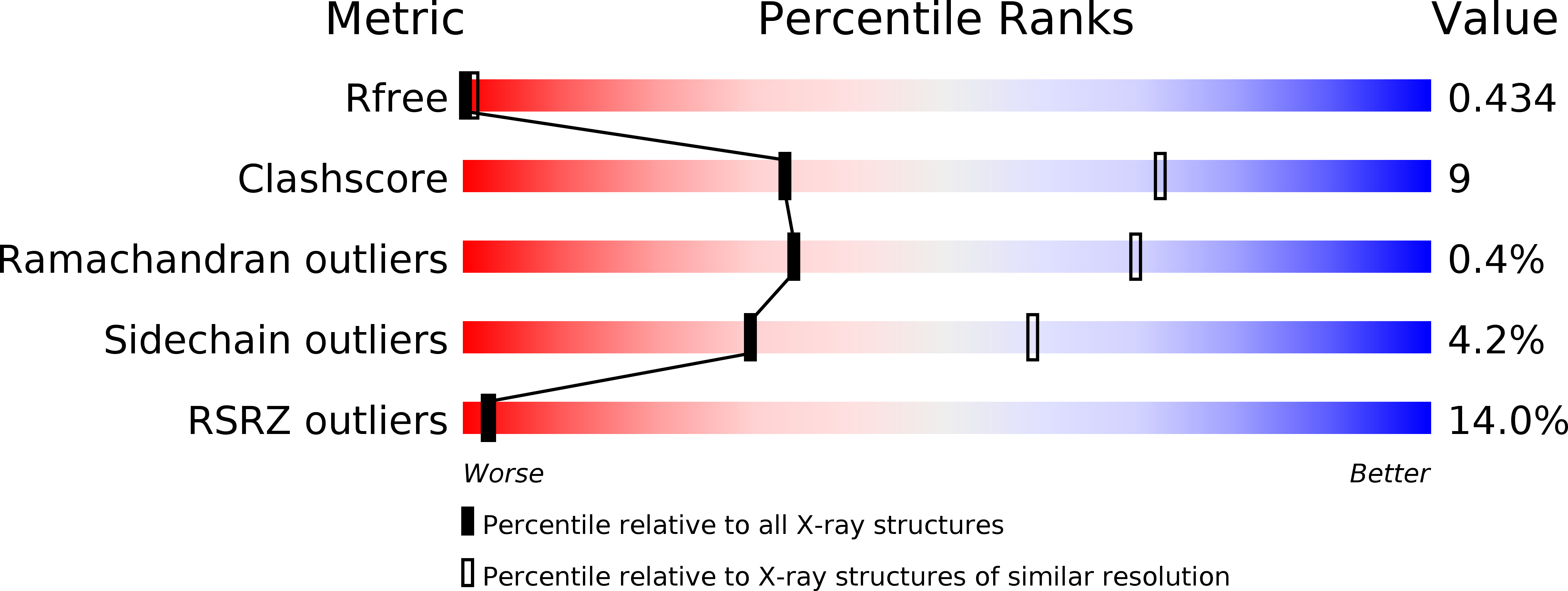
Resolution:
3.71 Å
R-Value Free:
0.43
R-Value Work:
0.39
R-Value Observed:
0.39
Space Group:
I 41 2 2
Macromolecular Entities
Structures with similar UniProt ID
Protein Blast
Polymer Type:polypeptide(L)
Molecule:Transposase
Gene (Uniprot):A
Mutagens:M521L, N525L
Chain IDs:A, B
Chain Length:529
Number of Molecules:2
Biological Source:Enterobacteria phage Mu
Polymer Type:polydeoxyribonucleotide
Molecule:DNA (68-MER)
Chain IDs:C
Chain Length:68
Number of Molecules:1
Biological Source:Enterobacteria phage Mu
Polymer Type:polydeoxyribonucleotide
Molecule:DNA (49-MER)
Chain IDs:E (auth: D)
Chain Length:49
Number of Molecules:1
Biological Source:Enterobacteria phage Mu
Polymer Type:polydeoxyribonucleotide
Molecule:DNA (13-MER)
Chain IDs:D (auth: E)
Chain Length:13
Number of Molecules:1
Biological Source:Enterobacteria phage Mu
Ligand Molecules
Primary Citation
The Mu transpososome structure sheds light on DDE recombinase evolution.
Nature 491 413 417 (2012)
PMID: 23135398 DOI: 10.1038/nature11602

Abstact

Studies of bacteriophage Mu transposition paved the way for understanding retroviral integration and V(D)J recombination as well as many other DNA transposition reactions. Here we report the structure of the Mu transpososome--Mu transposase (MuA) in complex with bacteriophage DNA ends and target DNA--determined from data that extend anisotropically to 5.2 Å, 5.2 Å and 3.7 Å resolution, in conjunction with previously determined structures of individual domains. The highly intertwined structure illustrates why chemical activity depends on formation of the synaptic complex, and reveals that individual domains have different roles when bound to different sites. The structure also provides explanations for the increased stability of the final product complex and for its preferential recognition by the ATP-dependent unfoldase ClpX. Although MuA and many other recombinases share a structurally conserved 'DDE' catalytic domain, comparisons among the limited set of available complex structures indicate that some conserved features, such as catalysis in trans and target DNA bending, arose through convergent evolution because they are important for function.

Legend

Protein

Chemical

Disease

Primary Citation of related structures