4FBZ image
Deposition Date 2012-05-23
Release Date 2013-05-15
Last Version Date 2024-10-30
Entry Detail
PDB ID:
4FBZ
Title:
Crystal structure of deltarhodopsin from Haloterrigena thermotolerans
Biological Source:
Source Organism:
Method Details:
Experimental Method:
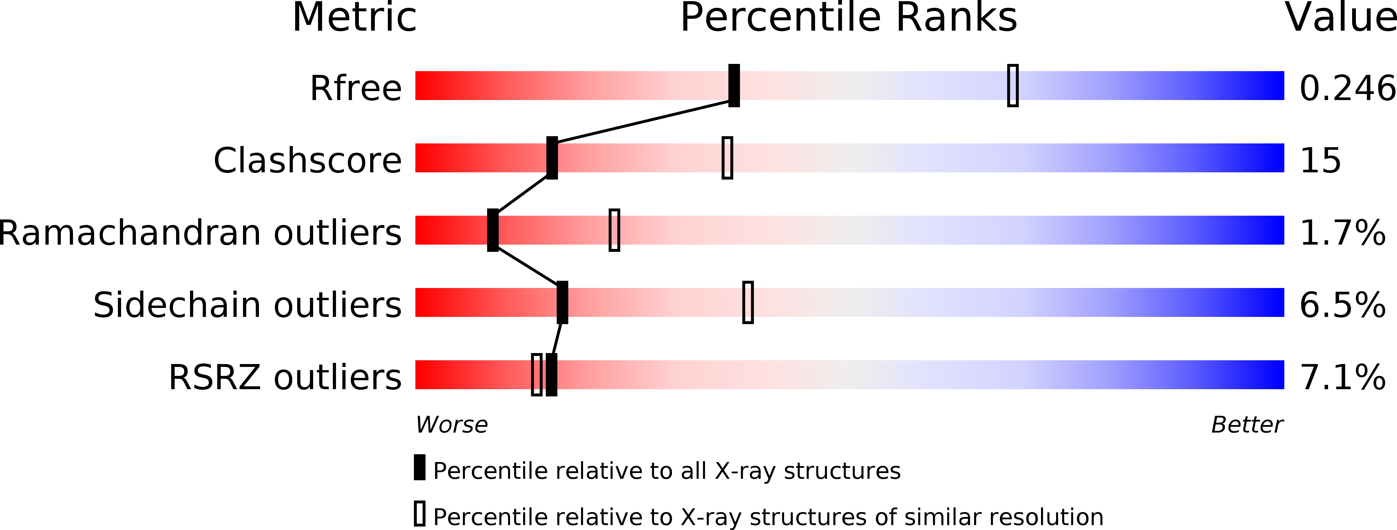
Resolution:
2.70 Å
R-Value Free:
0.26
R-Value Work:
0.23
R-Value Observed:
0.23
Space Group:
H 3 2
Macromolecular Entities
Structures with similar UniProt ID
Protein Blast
Polymer Type:polypeptide(L)
Molecule:deltarhodopsin
Chain IDs:A
Chain Length:241
Number of Molecules:1
Biological Source:Haloterrigena thermotolerans
Primary Citation
Crystal structure of deltarhodopsin-3 from Haloterrigena thermotolerans
Proteins 81 1585 1592 (2013)
PMID: 23625688 DOI: 10.1002/prot.24316

Abstact

Deltarhodopsin, a new member of the microbial rhodopsin family, functions as a light-driven proton pump. Here, we report the three-dimensional structure of deltarhodopsin (dR3) from Haloterrigena thermotolerans at 2.7 Å resolution. A crystal belonging to space group R32 (a, b = 111.71 Å, c = 198.25 Å) was obtained by the membrane fusion method. In this crystal, dR3 forms a trimeric structure as observed for bacteriorhodopsin (bR). Structural comparison of dR with bR showed that the inner part (the proton release and uptake pathways) is highly conserved. Meanwhile, residues in the protein-protein contact region are largely altered so that the diameter of the trimeric structure at the cytoplasmic side is noticeably larger in dR3. Unlike bR, dR3 possesses a helical segment at the C-terminal region that fills the space between the AB and EF loops. A significant difference is also seen in the FG loop, which is one residue longer in dR3. Another peculiar property of dR3 is a highly crowded distribution of positively charged residues on the cytoplasmic surface, which may be relevant to a specific interaction with some cytoplasmic component.

Legend

Protein

Chemical

Disease

Primary Citation of related structures
Feedback Form
Name
Email
Institute
Feedback