4F52 image
Deposition Date 2012-05-11
Release Date 2012-09-19
Last Version Date 2024-02-28
Entry Detail
PDB ID:
4F52
Title:
Structure of a Glomulin-RBX1-CUL1 complex
Biological Source:
Source Organism:
Homo sapiens (Taxon ID: 9606)
Host Organism:
Method Details:
Experimental Method:
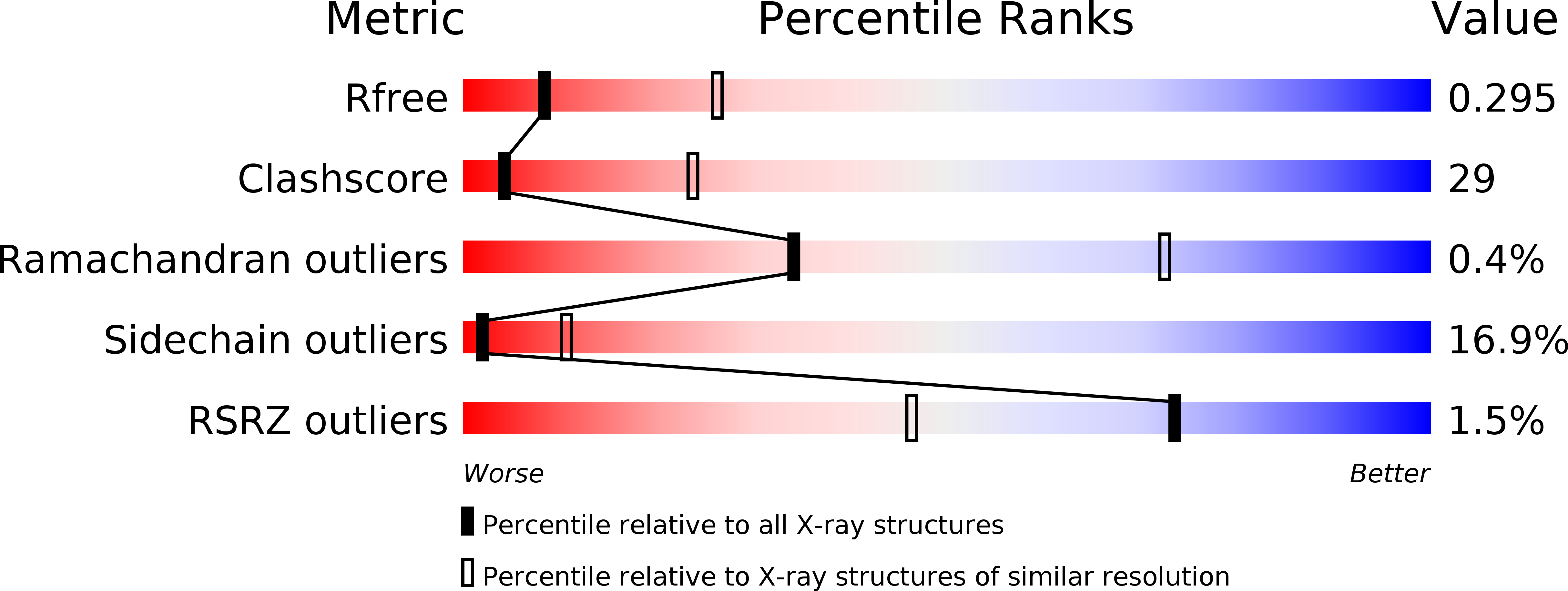
Resolution:
3.00 Å
R-Value Free:
0.28
R-Value Work:
0.21
R-Value Observed:
0.22
Space Group:
P 1 21 1
Macromolecular Entities
Polymer Type:polypeptide(L)
Molecule:Cullin-1
Gene (Uniprot):CUL1
Mutations:L421E, V451E, V452K, Y455K
Chain IDs:A, C
Chain Length:282
Number of Molecules:2
Biological Source:Homo sapiens
Polymer Type:polypeptide(L)
Molecule:E3 ubiquitin-protein ligase RBX1
Gene (Uniprot):RBX1
Chain IDs:B, D
Chain Length:106
Number of Molecules:2
Biological Source:Homo sapiens
Polymer Type:polypeptide(L)
Molecule:Glomulin
Gene (Uniprot):GLMN
Chain IDs:E, F
Chain Length:596
Number of Molecules:2
Biological Source:Homo sapiens
Ligand Molecules
Primary Citation
Structure of a Glomulin-RBX1-CUL1 Complex: Inhibition of a RING E3 Ligase through Masking of Its E2-Binding Surface.
Mol.Cell 47 371 382 (2012)
PMID: 22748924 DOI: 10.1016/j.molcel.2012.05.044

Abstact

The approximately 300 human cullin-RING ligases (CRLs) are multisubunit E3s in which a RING protein, either RBX1 or RBX2, recruits an E2 to catalyze ubiquitination. RBX1-containing CRLs also can bind Glomulin (GLMN), which binds RBX1's RING domain, regulates the RBX1-CUL1-containing SCF(FBW7) complex, and is disrupted in the disease Glomuvenous Malformation. Here we report the crystal structure of a complex between GLMN, RBX1, and a fragment of CUL1. Structural and biochemical analyses reveal that GLMN adopts a HEAT-like repeat fold that tightly binds the E2-interacting surface of RBX1, inhibiting CRL-mediated chain formation by the E2 CDC34. The structure explains the basis for GLMN's selectivity toward RBX1 over RBX2, and how disease-associated mutations disrupt GLMN-RBX1 interactions. Our study reveals a mechanism for RING E3 ligase regulation, whereby an inhibitor blocks E2 access, and raises the possibility that other E3s are likewise controlled by cellular proteins that mask E2-binding surfaces to mediate inhibition.

Legend

Protein

Chemical

Disease

Primary Citation of related structures