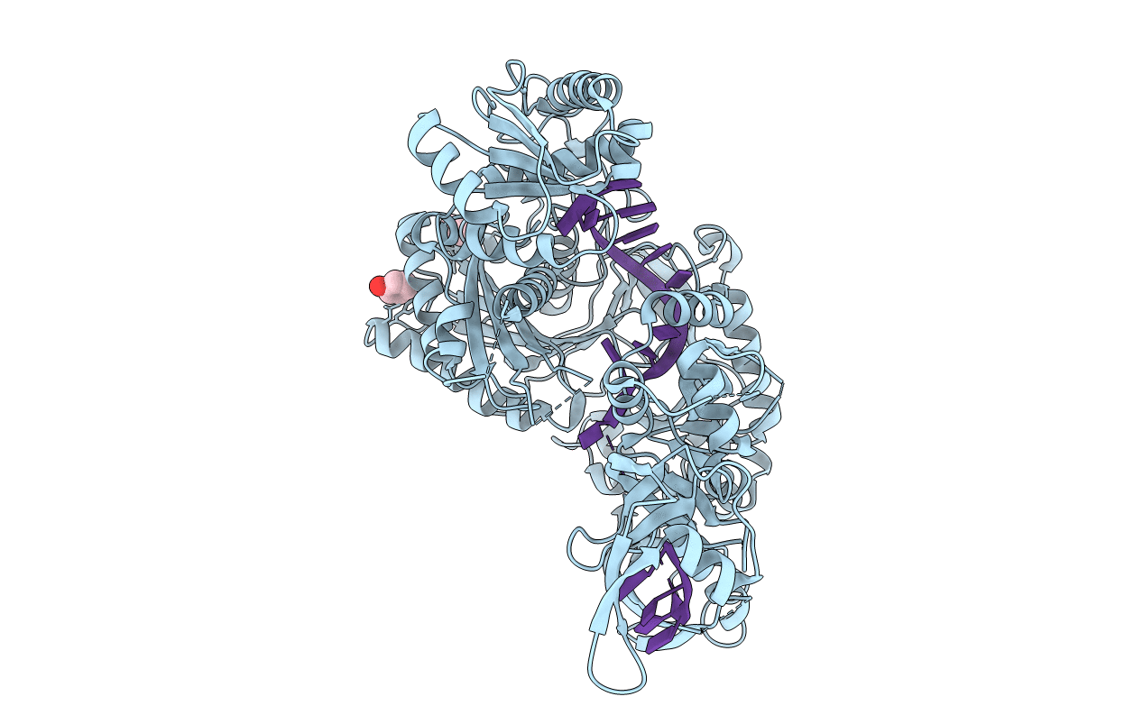
4F3T image
Entry Detail
PDB ID:
4F3T
Keywords:
Title:
Human Argonaute-2 - miR-20a complex
Biological Source:
Source Organism:
Host Organism:
PDB Version:
Deposition Date:
2012-05-09
Release Date:
2012-05-30
Method Details:
Experimental Method:
Resolution:
2.25 Å
R-Value Free:
0.25
R-Value Work:
0.20
R-Value Observed:
0.20
Space Group:
P 1 21 1
Macromolecular Entities
Polymer Type:polypeptide(L)
Description:Protein argonaute-2
Chain IDs:A
Chain Length:861
Number of Molecules:1
Biological Source:Homo sapiens
Polymer Type:polyribonucleotide
Description:RNA (5'-R(P*UP*AP*AP*AP*GP*UP*GP*CP*UP*UP*AP*UP*AP*GP*UP*G*CP*AP*GP*G)-3')
Chain IDs:B (auth: R)
Chain Length:20
Number of Molecules:1
Biological Source:Homo sapiens
Ligand Molecules
Primary Citation
The Structure of Human Argonaute-2 in Complex with miR-20a.
Cell(Cambridge,Mass.) 150 100 110 (2012)
PMID: 22682761 DOI: 10.1016/j.cell.2012.05.017

Abstact

Argonaute proteins lie at the heart of the RNA-induced silencing complex (RISC), wherein they use small RNA guides to recognize targets. Initial insight into the architecture of Argonautes came from studies of prokaryotic proteins, revealing a crescent-shaped base made up of the amino-terminal, PAZ, middle, and PIWI domains. The recently reported crystal structure of human Argonaute-2 (hAgo2), the "slicer" in RNA interference, in complex with a mixed population of RNAs derived from insect cells provides insight into the architecture of a eukaryotic Argonaute protein with defined biochemical and biological functions. Here, we report the structure of human Ago2 bound to a physiologically relevant microRNA, microRNA-20a, at 2.2 Å resolution. The miRNA is anchored at both ends by the Mid and PAZ domains and makes several kinks and turns along the binding groove. Interestingly, miRNA binding confers remarkable stability on hAgo2, locking this otherwise flexible enzyme into a stable conformation.

Legend

Protein

Chemical

Disease

Primary Citation of related structures