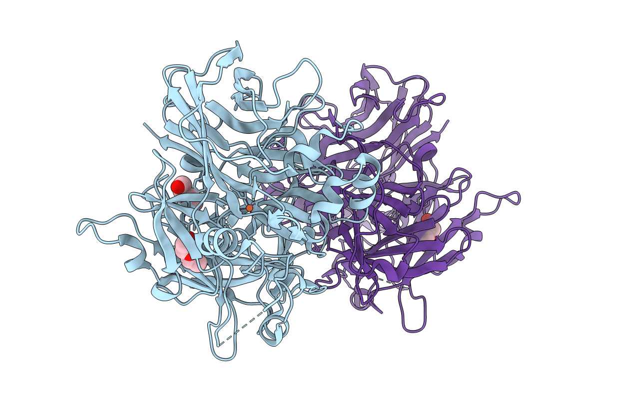
4F3D image
Deposition Date 2012-05-09
Release Date 2012-10-03
Last Version Date 2023-09-13
Entry Detail
PDB ID:
4F3D
Title:
Structure of RPE65: P65 crystal form grown in Fos-Choline-10
Biological Source:
Source Organism:
Bos taurus (Taxon ID: 9913)
Method Details:
Experimental Method:
Resolution:
2.50 Å
R-Value Free:
0.21
R-Value Work:
0.19
R-Value Observed:
0.19
Space Group:
P 65
Macromolecular Entities
Polymer Type:polypeptide(L)
Molecule:Retinoid isomerohydrolase
Gene (Uniprot):RPE65
Chain IDs:A, B
Chain Length:533
Number of Molecules:2
Biological Source:Bos taurus
Primary Citation
Structure of RPE65 isomerase in a lipidic matrix reveals roles for phospholipids and iron in catalysis.
Proc.Natl.Acad.Sci.USA 109 E2747 E2756 (2012)
PMID: 23012475 DOI: 10.1073/pnas.1212025109

Abstact

RPE65 is a key metalloenzyme responsible for maintaining visual function in vertebrates. Despite extensive research on this membrane-bound retinoid isomerase, fundamental questions regarding its enzymology remain unanswered. Here, we report the crystal structure of RPE65 in a membrane-like environment. These crystals, obtained from enzymatically active, nondelipidated protein, displayed an unusual packing arrangement wherein RPE65 is embedded in a lipid-detergent sheet. Structural differences between delipidated and nondelipidated RPE65 uncovered key residues involved in substrate uptake and processing. Complementary iron K-edge X-ray absorption spectroscopy data established that RPE65 as isolated contained a divalent iron center and demonstrated the presence of a tightly bound ligand consistent with a coordinated carboxylate group. These results support the hypothesis that the Lewis acidity of iron could be used to promote ester dissociation and generation of a carbocation intermediate required for retinoid isomerization.

Legend

Protein

Chemical

Disease

Primary Citation of related structures