
Deposition Date
2012-05-08
Release Date
2012-10-03
Last Version Date
2023-09-13
Entry Detail
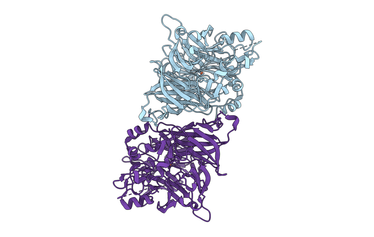
PDB ID:
4F2Z
Keywords:
Title:
Crystal structure of RPE65 in a lipid environment
Biological Source:
Source Organism(s):
Bos taurus (Taxon ID: 9913)
Method Details:
Experimental Method:
Resolution:
3.00 Å
R-Value Free:
0.26
R-Value Work:
0.22
R-Value Observed:
0.22
Space Group:
P 21 21 21
