4F2M image
Entry Detail
PDB ID:
4F2M
Title:
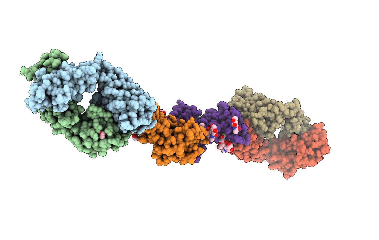
Crystal structure of a TGEV coronavirus Spike fragment in complex with the TGEV neutralizing monoclonal antibody 1AF10
Biological Source:
PDB Version:
Deposition Date:
2012-05-08
Release Date:
2012-08-22
Method Details:
Experimental Method:
Resolution:
3.00 Å
R-Value Free:
0.25
R-Value Work:
0.21
R-Value Observed:
0.21
Space Group:
P 1 21 1
Macromolecular Entities
Polymer Type:polypeptide(L)
Description:monoclonal antibody 1AF10, heavy chain
Chain IDs:A, C
Chain Length:221
Number of Molecules:2
Biological Source:Mus musculus
Polymer Type:polypeptide(L)
Description:monoclonal antibody 1AF10, light chain
Chain IDs:B, D
Chain Length:214
Number of Molecules:2
Biological Source:Mus musculus
Polymer Type:polypeptide(L)
Description:Spike protein
Chain IDs:E, F
Chain Length:209
Number of Molecules:2
Biological Source:TGEV virulent Purdue
Modified Residue
Compound ID Chain ID Parent Comp ID Details 2D Image
ASN E ASN GLYCOSYLATION SITE
Primary Citation
Structural bases of coronavirus attachment to host aminopeptidase N and its inhibition by neutralizing antibodies.
Plos Pathog. 8 e1002859 e1002859 (2012)
PMID: 22876187 DOI: 10.1371/journal.ppat.1002859

Abstact

The coronaviruses (CoVs) are enveloped viruses of animals and humans associated mostly with enteric and respiratory diseases, such as the severe acute respiratory syndrome and 10-20% of all common colds. A subset of CoVs uses the cell surface aminopeptidase N (APN), a membrane-bound metalloprotease, as a cell entry receptor. In these viruses, the envelope spike glycoprotein (S) mediates the attachment of the virus particles to APN and subsequent cell entry, which can be blocked by neutralizing antibodies. Here we describe the crystal structures of the receptor-binding domains (RBDs) of two closely related CoV strains, transmissible gastroenteritis virus (TGEV) and porcine respiratory CoV (PRCV), in complex with their receptor, porcine APN (pAPN), or with a neutralizing antibody. The data provide detailed information on the architecture of the dimeric pAPN ectodomain and its interaction with the CoV S. We show that a protruding receptor-binding edge in the S determines virus-binding specificity for recessed glycan-containing surfaces in the membrane-distal region of the pAPN ectodomain. Comparison of the RBDs of TGEV and PRCV to those of other related CoVs, suggests that the conformation of the S receptor-binding region determines cell entry receptor specificity. Moreover, the receptor-binding edge is a major antigenic determinant in the TGEV envelope S that is targeted by neutralizing antibodies. Our results provide a compelling view on CoV cell entry and immune neutralization, and may aid the design of antivirals or CoV vaccines. APN is also considered a target for cancer therapy and its structure, reported here, could facilitate the development of anti-cancer drugs.

Legend

Protein

Chemical

Disease

Primary Citation of related structures