4F2I image
Deposition Date 2012-05-08
Release Date 2013-05-08
Last Version Date 2024-10-30
Entry Detail
PDB ID:
4F2I
Title:
Crystal structure of glutaredoxin-like NrdH from Mycobacterium tuberculosis
Biological Source:
Source Organism:
Host Organism:
Method Details:
Experimental Method:
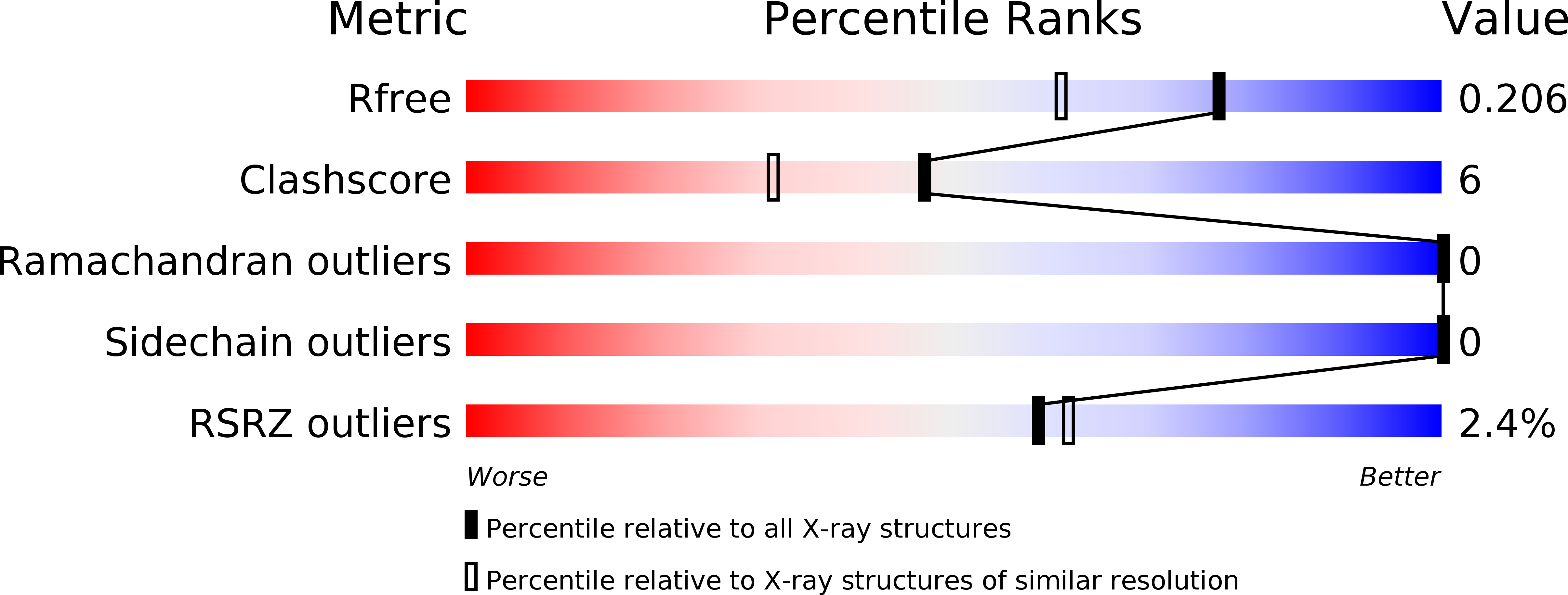
Resolution:
1.67 Å
R-Value Free:
0.21
R-Value Work:
0.17
R-Value Observed:
0.17
Space Group:
P 21 21 2
Macromolecular Entities
Structures with similar UniProt ID
Protein Blast
Polymer Type:polypeptide(L)
Molecule:Glutaredoxin NrdH, putative
Chain IDs:A
Chain Length:84
Number of Molecules:1
Biological Source:Mycobacterium tuberculosis
Ligand Molecules
Primary Citation
The Crystal Structure of Mycobacterium tuberculosis NrdH at 0.87 A Suggests a Possible Mode of Its Activity.
Biochemistry 52 4056 4065 (2013)
PMID: 23675692 DOI: 10.1021/bi400191z

Abstact

Members of the NrdH family of redox proteins, which consists of small glutaredoxin-like proteins with thioredoxin-like activity, serve as the reducing partners of class Ib ribonucleotide reductases. Here, we report the crystal structure of NrdH from Mycobacterium tuberculosis, refined to a crystallographic R factor of 14.02% (Rfree = 15.53%) at 0.87 Å resolution. The tertiary structure of M. tuberculosis NrdH has a typical thioredoxin fold as expected. The extremely high resolution of the structure allows us to dissect the functionality of the protein in great depth. Structural superimposition of M. tuberculosis NrdH and thioredoxin reductase over the Escherichia coli thioredoxin reductase-thioredoxin complex suggests the ability of NrdH to accept electrons from M. tuberculosis thioredoxin reductase. This raises the important question of why glutaredoxins are unable to accept electrons from thioredoxin reductases and why thioredoxins are unable to reduce ribonucleotide reductases. Furthermore, forms of NrdH from other organisms have been shown to be a specific reductant of class Ib ribonucleotide reductases. We attempt to explain this substrate specificity by modeling the C-terminal peptide of a ribunucleotide subunit, NrdE, in the active site of NrdH using the already available Grx-NrdA-Cter-peptide structure. Statistical coupling analysis of NrdH, glutaredoxins, and thioredoxins reveals different sets of co-evolving contiguous clusters of amino acid residues, which might explain the differences in the biochemical properties of these structurally similar yet functionally distinct subclasses of proteins.

Legend

Protein

Chemical

Disease

Primary Citation of related structures