
Deposition Date
2012-05-05
Release Date
2012-06-06
Last Version Date
2024-10-30
Entry Detail
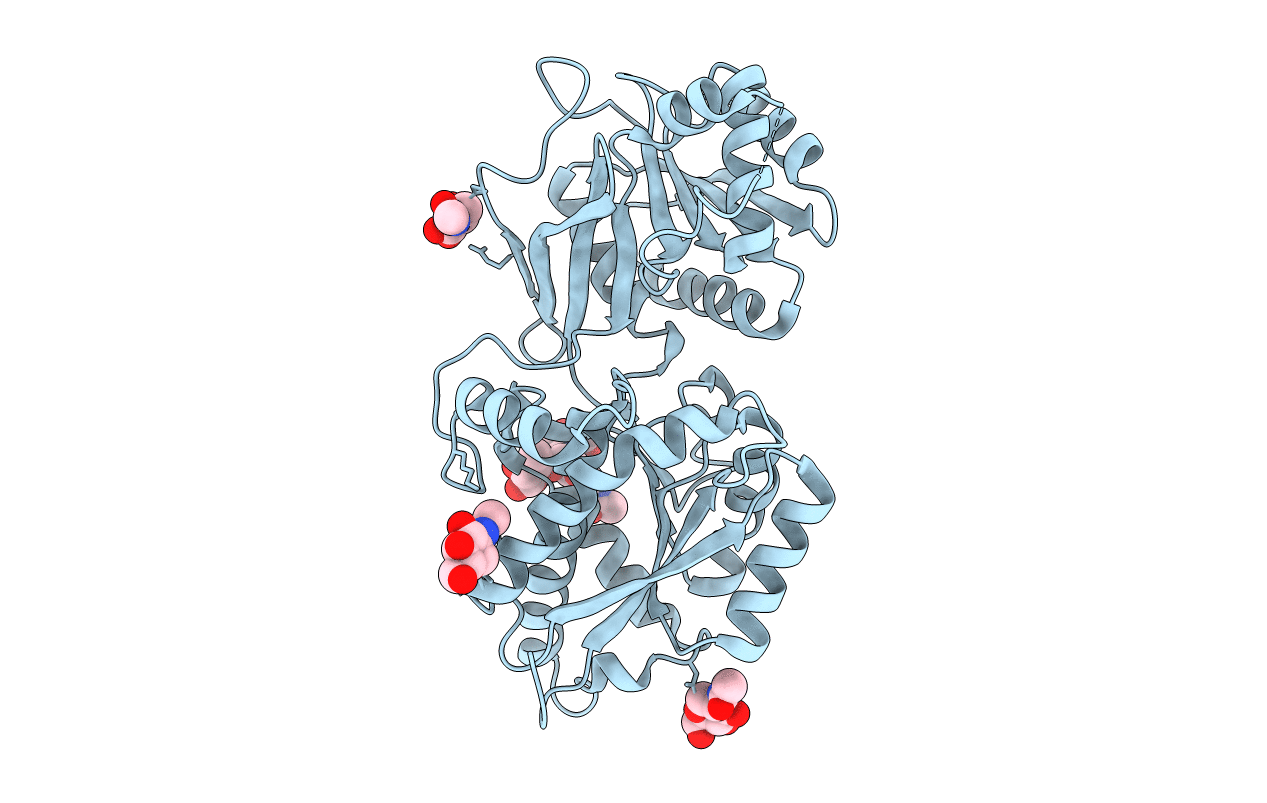
PDB ID:
4F12
Keywords:
Title:
Crystal structure of the extracellular domain of human GABA(B) receptor GBR2
Biological Source:
Source Organism(s):
Homo sapiens (Taxon ID: 9606)
Expression System(s):
Method Details:
Experimental Method:
Resolution:
3.02 Å
R-Value Free:
0.25
R-Value Work:
0.20
R-Value Observed:
0.20
Space Group:
C 2 2 21
