4EZD image
Entry Detail
PDB ID:
4EZD
Title:
Crystal Structure of the UT-B Urea Transporter from Bos Taurus Bound to Selenourea
Biological Source:
Source Organism:
Host Organism:
PDB Version:
Deposition Date:
2012-05-02
Release Date:
2012-06-27
Method Details:
Experimental Method:
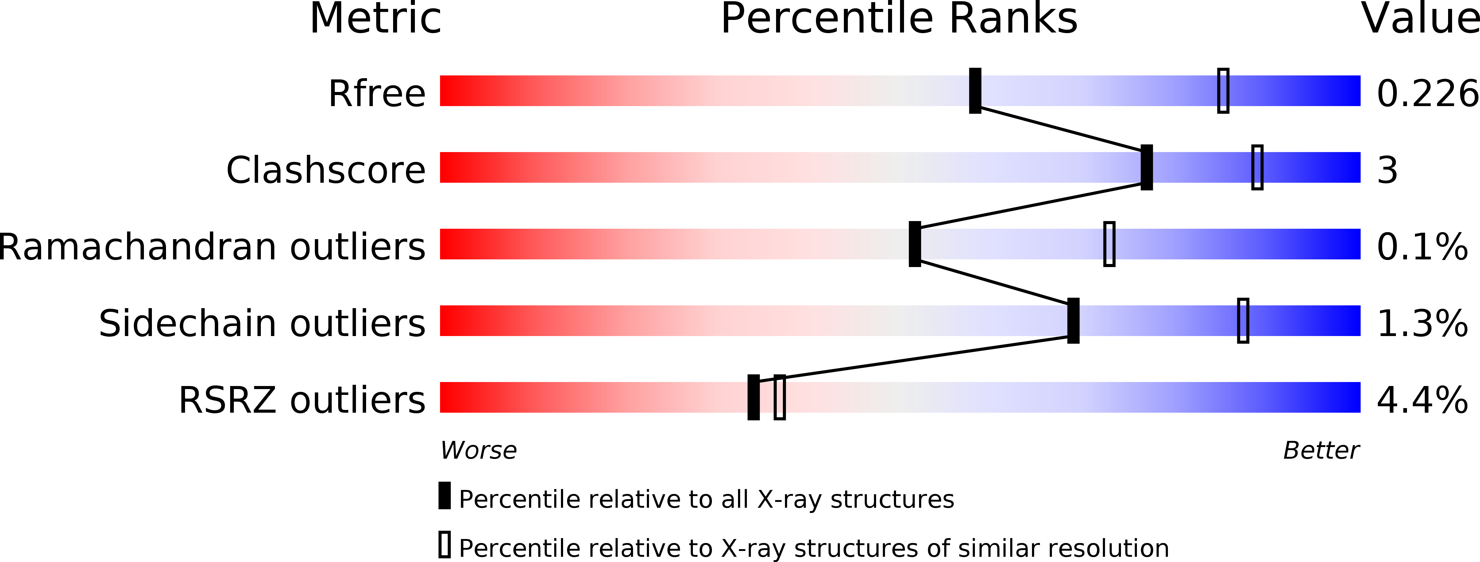
Resolution:
2.50 Å
R-Value Free:
0.22
R-Value Work:
0.19
R-Value Observed:
0.19
Space Group:
P 1 21 1
Macromolecular Entities
Polymer Type:polypeptide(L)
Description:Urea transporter 1
Chain IDs:A, B, C
Chain Length:384
Number of Molecules:3
Biological Source:Bos taurus
Primary Citation
Structure and permeation mechanism of a mammalian urea transporter.
Proc.Natl.Acad.Sci.USA 109 11194 11199 (2012)
PMID: 22733730 DOI: 10.1073/pnas.1207362109

Abstact

As an adaptation to infrequent access to water, terrestrial mammals produce urine that is hyperosmotic to plasma. To prevent osmotic diuresis by the large quantity of urea generated by protein catabolism, the kidney epithelia contain facilitative urea transporters (UTs) that allow rapid equilibration between the urinary space and the hyperosmotic interstitium. Here we report the first X-ray crystal structure of a mammalian UT, UT-B, at a resolution of 2.36 Å. UT-B is a homotrimer and each protomer contains a urea conduction pore with a narrow selectivity filter. Structural analyses and molecular dynamics simulations showed that the selectivity filter has two urea binding sites separated by an approximately 5.0 kcal/mol energy barrier. Functional studies showed that the rate of urea conduction in UT-B is increased by hypoosmotic stress, and that the site of osmoregulation coincides with the location of the energy barrier.

Legend

Protein

Chemical

Disease

Primary Citation of related structures