Deposition Date
2012-04-30
Release Date
2012-05-30
Last Version Date
2024-11-06
Entry Detail
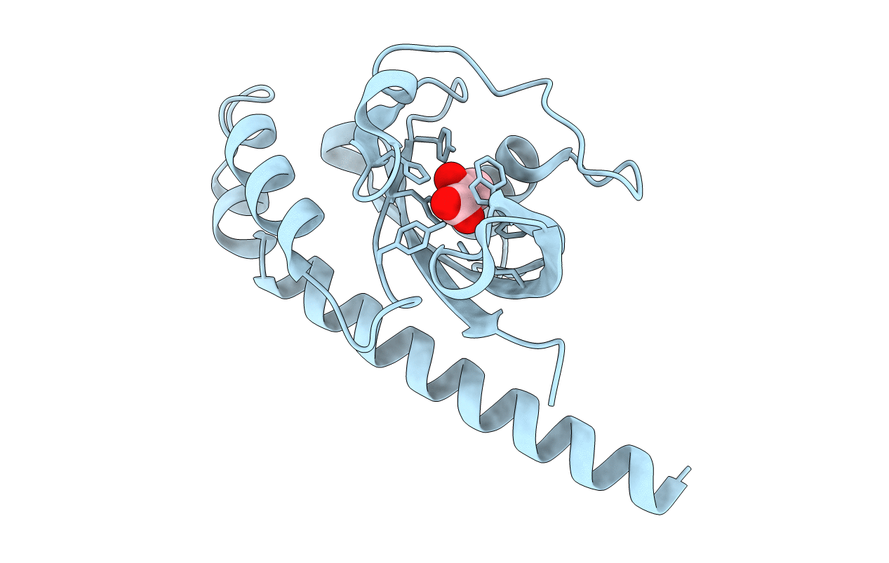
PDB ID:
4EXO
Keywords:
Title:
Revised, rerefined crystal structure of PDB entry 2QHK, methyl accepting chemotaxis protein
Biological Source:
Source Organism(s):
Vibrio parahaemolyticus (Taxon ID: 670)
Expression System(s):
Method Details:
Experimental Method:
Resolution:
1.90 Å
R-Value Free:
0.20
R-Value Work:
0.15
R-Value Observed:
0.15
Space Group:
I 4 2 2