Deposition Date
2012-04-23
Release Date
2013-05-08
Last Version Date
2023-09-13
Entry Detail
PDB ID:
4ESM
Keywords:
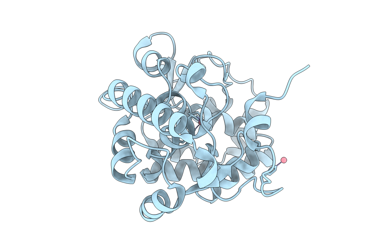
Title:
Crystallographic structure of phenylalanine hydroxylase from Chromobacterium violaceum Y155A mutation
Biological Source:
Source Organism(s):
Chromobacterium violaceum (Taxon ID: 243365)
Expression System(s):
Method Details:
Experimental Method:
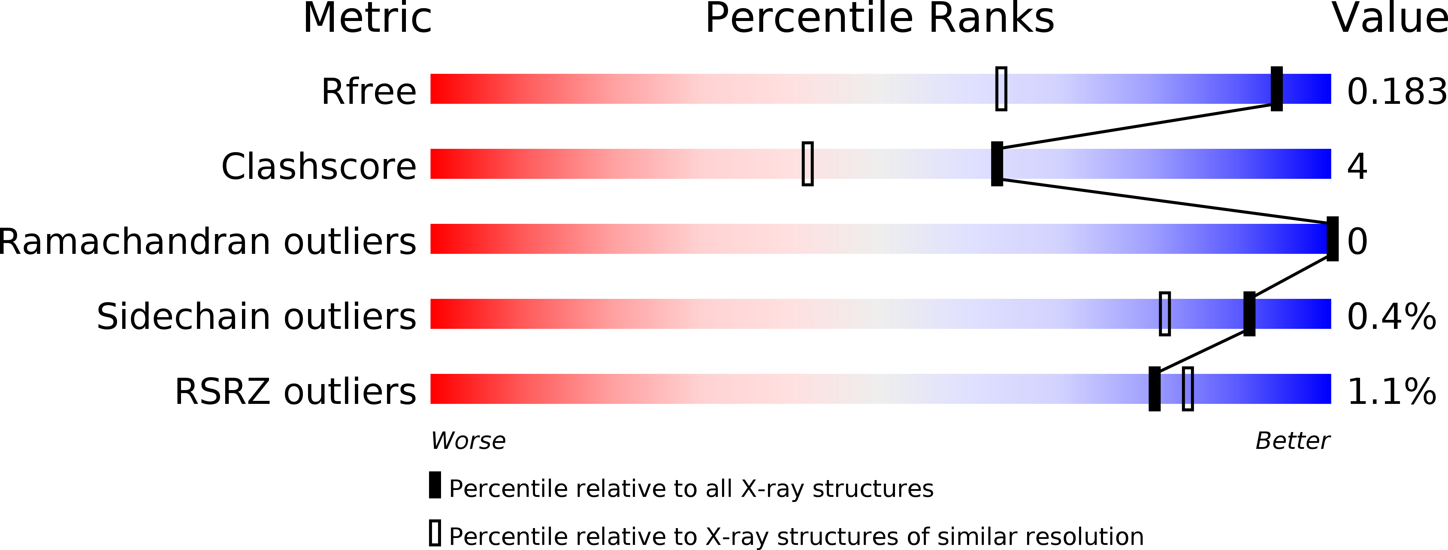
Resolution:
1.35 Å
R-Value Free:
0.18
R-Value Work:
0.15
R-Value Observed:
0.15
Space Group:
P 1