4ERM image
Entry Detail
PDB ID:
4ERM
Keywords:
Title:
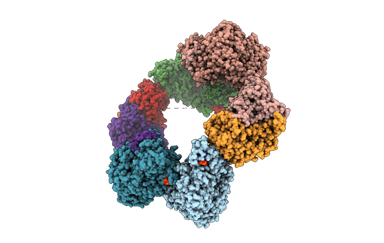
Crystal structure of the dATP inhibited E. coli class Ia ribonucleotide reductase complex at 4 Angstroms resolution
Biological Source:
Source Organism:
Host Organism:
PDB Version:
Deposition Date:
2012-04-20
Release Date:
2012-07-04
Method Details:
Experimental Method:
Resolution:
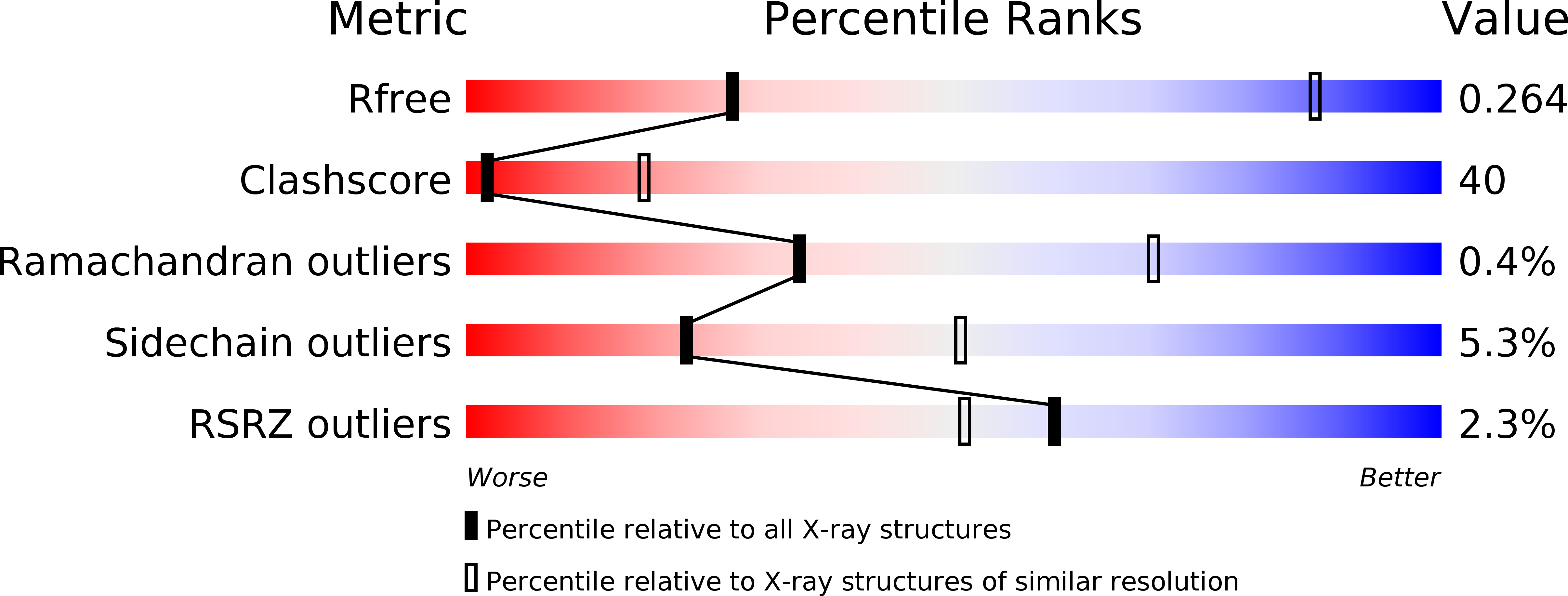
3.95 Å
R-Value Free:
0.28
R-Value Work:
0.25
Space Group:
C 1 2 1
Macromolecular Entities
Polymer Type:polypeptide(L)
Description:Ribonucleoside-diphosphate reductase 1 subunit alpha
Chain IDs:A, B, C, D
Chain Length:761
Number of Molecules:4
Biological Source:Escherichia coli K-12
Polymer Type:polypeptide(L)
Description:Ribonucleoside-diphosphate reductase 1 subunit beta
Chain IDs:E, F, G, H
Chain Length:375
Number of Molecules:4
Biological Source:Escherichia coli K-12
Primary Citation
Tangled up in knots: structures of inactivated forms of E. coli class Ia ribonucleotide reductase.
Structure 20 1374 1383 (2012)
PMID: 22727814 DOI: 10.1016/j.str.2012.05.009

Abstact

Ribonucleotide reductases (RNRs) provide the precursors for DNA biosynthesis and repair and are successful targets for anticancer drugs such as clofarabine and gemcitabine. Recently, we reported that dATP inhibits E. coli class Ia RNR by driving formation of RNR subunits into α4β4 rings. Here, we present the first X-ray structure of a gemcitabine-inhibited E. coli RNR and show that the previously described α4β4 rings can interlock to form an unprecedented (α4β4)2 megacomplex. This complex is also seen in a higher-resolution dATP-inhibited RNR structure presented here, which employs a distinct crystal lattice from that observed in the gemcitabine-inhibited case. With few reported examples of protein catenanes, we use data from small-angle X-ray scattering and electron microscopy to both understand the solution conditions that contribute to concatenation in RNRs as well as present a mechanism for the formation of these unusual structures.

Legend

Protein

Chemical

Disease

Primary Citation of related structures