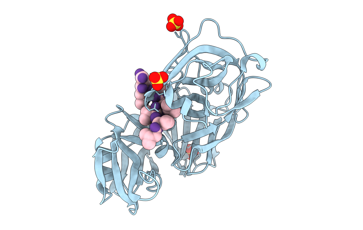
4ER2 image
Deposition Date 1990-10-20
Release Date 1991-01-15
Last Version Date 2024-10-30
Entry Detail
PDB ID:
4ER2
Title:
The active site of aspartic proteinases
Biological Source:
Method Details:
Experimental Method:
Resolution:
2.00 Å
R-Value Work:
0.18
Space Group:
P 1 21 1
Macromolecular Entities
Polymer Type:polypeptide(L)
Molecule:ENDOTHIAPEPSIN
Gene (Uniprot):EAPA
Chain IDs:A (auth: E)
Chain Length:330
Number of Molecules:1
Biological Source:Cryphonectria parasitica
Polymer Type:polypeptide(L)
Molecule:PEPSTATIN
Chain IDs:B (auth: I)
Chain Length:6
Number of Molecules:1
Biological Source:Streptomyces argenteolus subsp. toyonakensis
Ligand Molecules
Peptide-like Molecules
PRD_000557
Primary Citation

Abstact

The active site of the aspartic proteinase, endothiapepsin, has been defined by X-ray analysis and restrained least-squares refinement at 2.1 A resolution with a crystallographic agreement value of 0.16. The environments of the two catalytically important aspartyl groups are remarkably similar and the contributions of the NH2- and COOH-terminal domains to the catalytic centre are related by a local 2-fold axis. The carboxylates of the aspartyls share a hydrogen bond and have equivalent contacts to a bound water molecule or hydroxonium ion lying on the local diad. The main chains around 32 and 215 are connected by a novel interaction involving diad-related threonines. It is suggested that the two pKa values of the active site aspartyls arise from a structure not unlike that in maleic acid with a hydrogen-bonded intermediate species and a dicarboxylate characterised by electrostatic repulsions between the two negatively charged groups.

Legend

Protein

Chemical

Disease

Primary Citation of related structures