4EPC image
Deposition Date 2012-04-17
Release Date 2012-06-06
Last Version Date 2025-03-26
Entry Detail
PDB ID:
4EPC
Keywords:
Title:
Crystal structure of Autolysin repeat domains from Staphylococcus epidermidis
Biological Source:
Source Organism:
Host Organism:
Method Details:
Experimental Method:
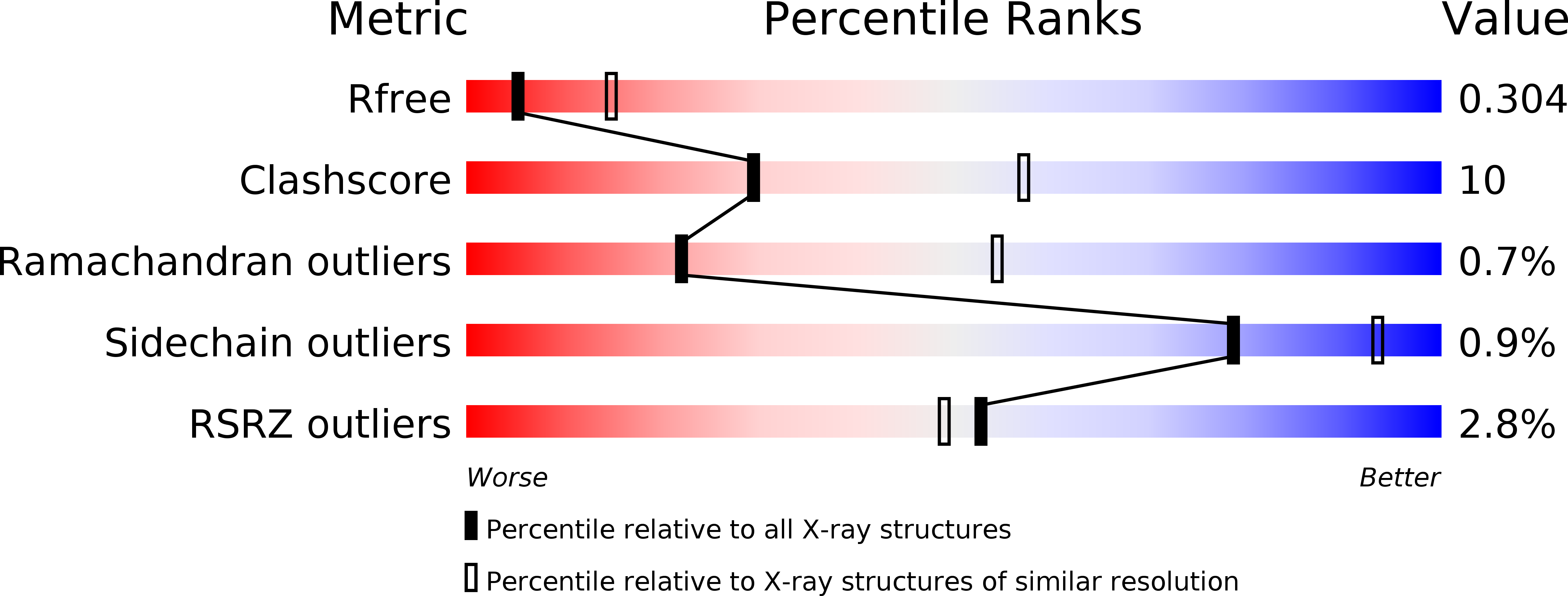
Resolution:
2.90 Å
R-Value Free:
0.29
R-Value Work:
0.26
R-Value Observed:
0.26
Space Group:
P 61 2 2
Macromolecular Entities
Structures with similar UniProt ID
Protein Blast
Polymer Type:polypeptide(L)
Molecule:N-acetylmuramoyl-L-alanine amidase
Gene (Uniprot):atl
Chain IDs:A
Chain Length:334
Number of Molecules:1
Biological Source:Staphylococcus epidermidis
Modified Residue
Compound ID Chain ID Parent Comp ID Details 2D Image
MLY A LYS N-DIMETHYL-LYSINE
Primary Citation
Ligand-binding properties and conformational dynamics of autolysin repeat domains in staphylococcal cell wall recognition.
J.Bacteriol. 194 3789 3802 (2012)
PMID: 22609916 DOI: 10.1128/JB.00331-12

Abstact

The bifunctional major autolysin Atl plays a key role in staphylococcal cell separation. Processing of Atl yields catalytically active amidase (AM) and glucosaminidase (GL) domains that are each fused to repeating units. The two repeats of AM (R1 and R2) target the enzyme to the septum, where it cleaves murein between dividing cells. We have determined the crystal structure of R2, which reveals that each repeat folds into two half-open β-barrel subunits. We further demonstrate that lipoteichoic acid serves as a receptor for the repeats and that this interaction depends on conserved surfaces in each subunit. Small-angle X-ray scattering of the mature amidase reveals the presence of flexible linkers separating the AM, R1, and R2 units. Different levels of flexibility for each linker provide mechanistic insights into the conformational dynamics of the full-length protein and the roles of its components in cell wall association and catalysis. Our analysis supports a model in which the repeats direct the catalytic AM domain to the septum, where it can optimally perform the final step of cell division.

Legend

Protein

Chemical

Disease

Primary Citation of related structures