Deposition Date
2012-04-15
Release Date
2012-04-25
Last Version Date
2024-11-06
Entry Detail
PDB ID:
4EOU
Keywords:
Title:
Crystal structure of E. coli dihydrodipicolinate synthase with pyruvate and succinic semi-aldehyde bound in active site
Biological Source:
Source Organism(s):
Escherichia coli (Taxon ID: 562)
Method Details:
Experimental Method:
Resolution:
2.30 Å
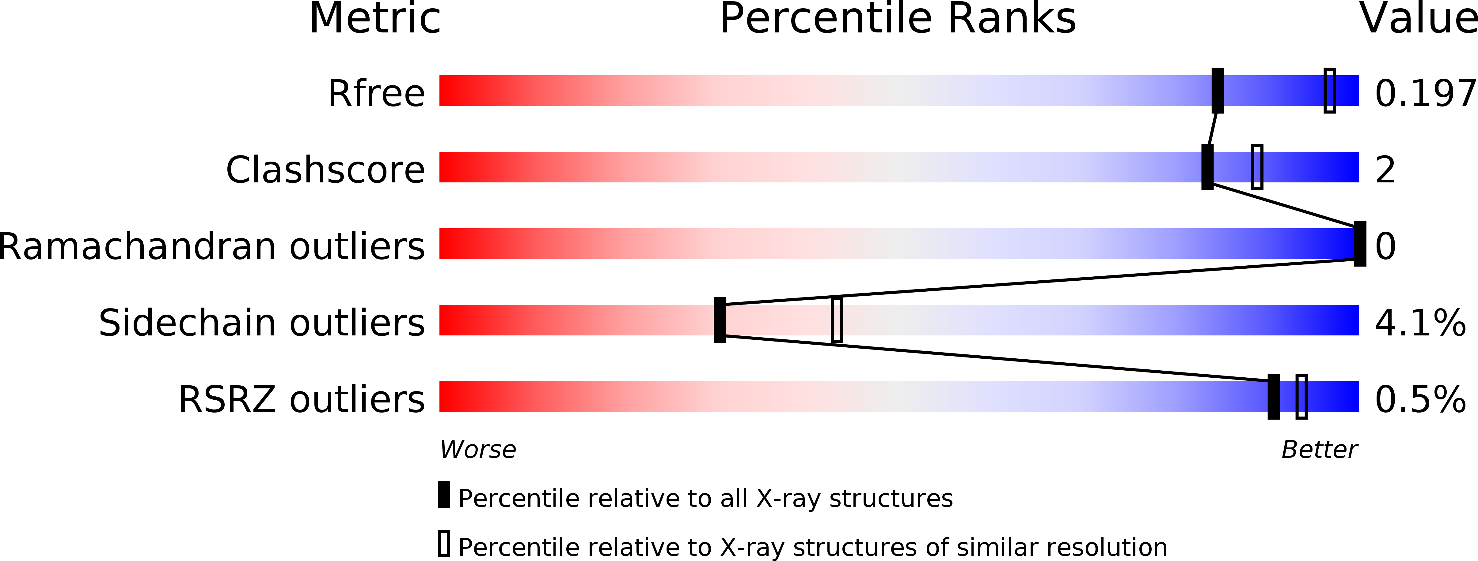
R-Value Free:
0.19
R-Value Work:
0.14
R-Value Observed:
0.14
Space Group:
P 31 2 1