4EOT image
Deposition Date 2012-04-15
Release Date 2012-11-28
Last Version Date 2023-09-13
Entry Detail
PDB ID:
4EOT
Title:
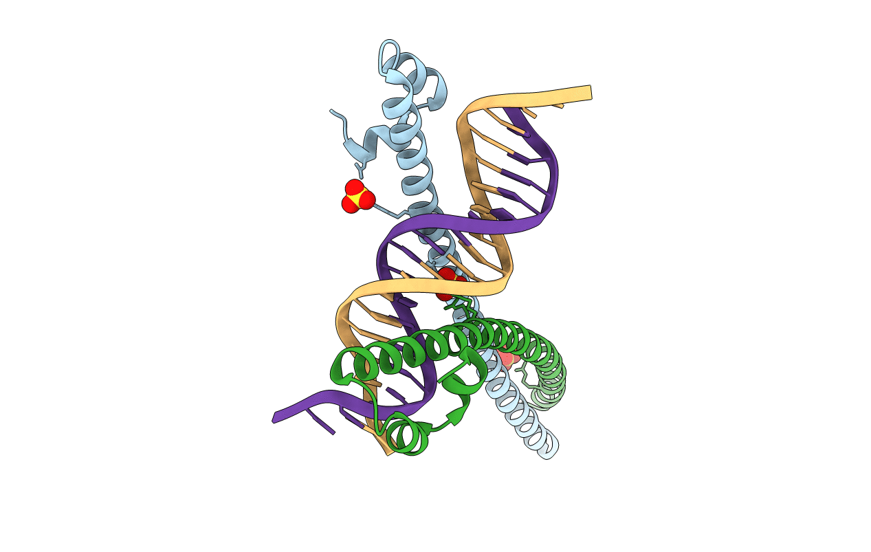
Crystal structure of the MafA homodimer bound to the consensus MARE
Biological Source:
Source Organism:
Homo sapiens (Taxon ID: 9606)
Host Organism:
Method Details:
Experimental Method:
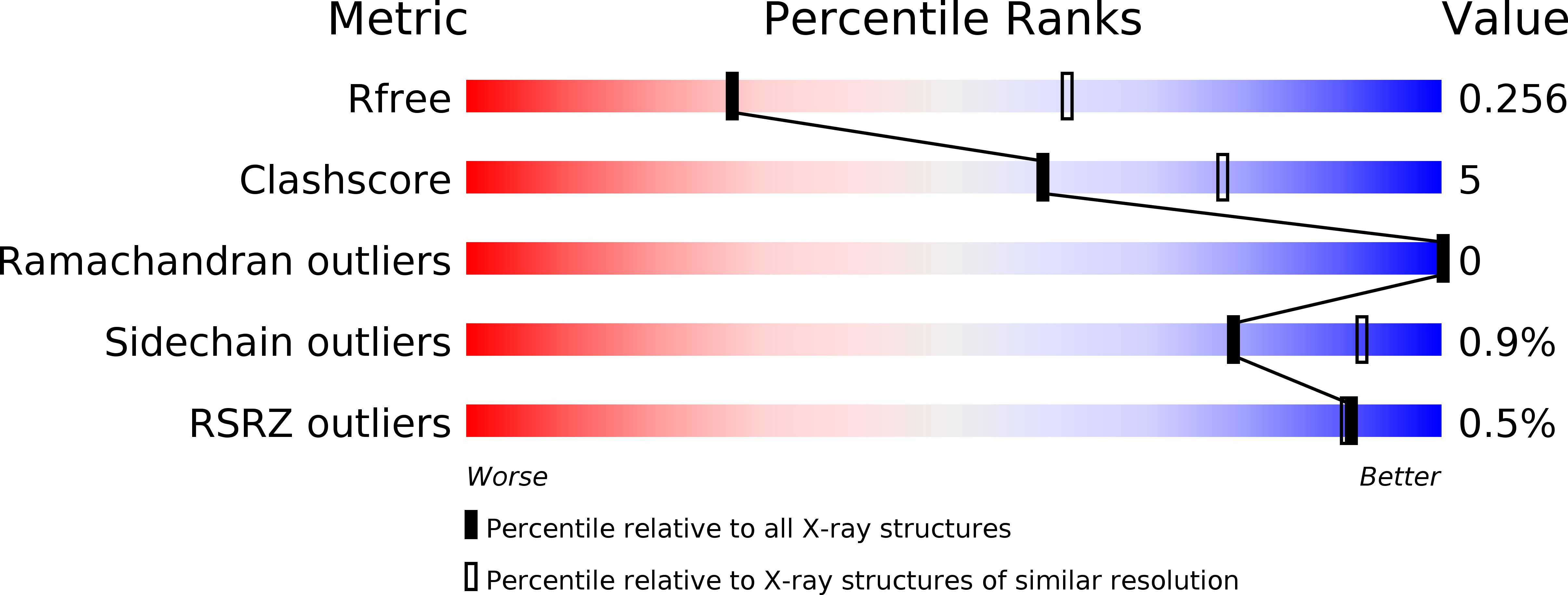
Resolution:
2.86 Å
R-Value Free:
0.25
R-Value Work:
0.22
R-Value Observed:
0.22
Space Group:
P 31 2 1
Macromolecular Entities
Structures with similar UniProt ID
Protein Blast
Polymer Type:polypeptide(L)
Molecule:Transcription factor MafA
Gene (Uniprot):MAFA
Chain IDs:A, B
Chain Length:95
Number of Molecules:2
Biological Source:Homo sapiens
Ligand Molecules
Primary Citation
A Novel DNA Binding Mechanism for maf Basic Region-Leucine Zipper Factors Inferred from a MafA-DNA Complex Structure and Binding Specificities.
Biochemistry 51 9706 9717 (2012)
PMID: 23148532 DOI: 10.1021/bi301248j

Abstact

MafA is a proto-oncoprotein and is critical for insulin gene expression in pancreatic β-cells. Maf proteins belong to the AP1 superfamily of basic region-leucine zipper (bZIP) transcription factors. Residues in the basic helix and an ancillary N-terminal domain, the Extended Homology Region (EHR), endow maf proteins with unique DNA binding properties: binding a 13 bp consensus site consisting of a core AP1 site (TGACTCA) flanked by TGC sequences and binding DNA stably as monomers. To further characterize maf DNA binding, we determined the structure of a MafA-DNA complex. MafA forms base-specific hydrogen bonds with the flanking G(-5)C(-4) and central C(0)/G(0) bases, but not with the core-TGA bases. However, in vitro binding studies utilizing a pulse-chase electrophoretic mobility shift assay protocol revealed that mutating either the core-TGA or flanking-TGC bases dramatically increases the binding off rate. Comparing the known maf structures, we propose that DNA binding specificity results from positioning the basic helix through unique phosphate contacts. The EHR does not contact DNA directly but stabilizes DNA binding by contacting the basic helix. Collectively, these results suggest a novel multistep DNA binding process involving a conformational change from contacting the core-TGA to contacting the flanking-TGC bases.

Legend

Protein

Chemical

Disease

Primary Citation of related structures