4EO2 image
Deposition Date 2012-04-13
Release Date 2012-08-22
Last Version Date 2024-02-28
Entry Detail
PDB ID:
4EO2
Title:
Structure of the bacteriophage C1 tail knob protein, gp12
Biological Source:
Source Organism:
Host Organism:
Method Details:
Experimental Method:
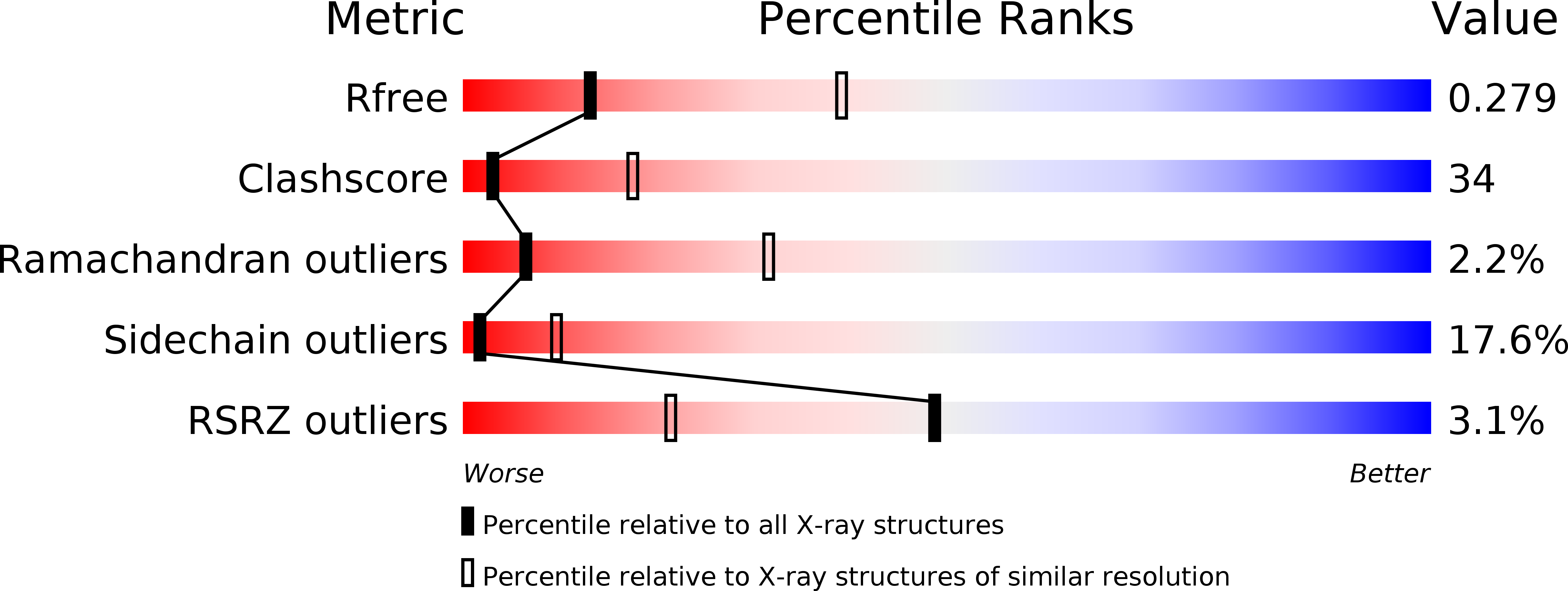
Resolution:
3.01 Å
R-Value Free:
0.28
R-Value Work:
0.23
R-Value Observed:
0.24
Space Group:
P 21 21 2
Macromolecular Entities
Polymer Type:polypeptide(L)
Molecule:Major tail protein
Gene (Uniprot):orf12
Chain IDs:A, B, C, D, E, F
Chain Length:583
Number of Molecules:6
Biological Source:Streptococcus phage C1
Ligand Molecules
Primary Citation
Structural investigations of a Podoviridae streptococcus phage C1, implications for the mechanism of viral entry.
Proc.Natl.Acad.Sci.USA 109 14001 14006 (2012)
PMID: 22891295 DOI: 10.1073/pnas.1207730109

Abstact

The Podoviridae phage C1 was one of the earliest isolated bacteriophages and the first virus documented to be active against streptococci. The icosahedral and asymmetric reconstructions of the virus were calculated using cryo-electron microscopy. The capsid protein has an HK97 fold arranged into a T = 4 icosahedral lattice. The C1 tail is terminated with a ϕ29-like knob, surrounded by a skirt of twelve long appendages with novel morphology. Several C1 structural proteins have been identified, including a candidate for an appendage. The crystal structure of the knob has an N-terminal domain with a fold observed previously in tube forming proteins of Siphoviridae and Myoviridae phages. The structure of C1 suggests the mechanisms by which the virus digests the cell wall and ejects its genome. Although there is little sequence similarity to other phages, conservation of the structural proteins demonstrates a common origin of the head and tail, but more recent evolution of the appendages.

Legend

Protein

Chemical

Disease

Primary Citation of related structures