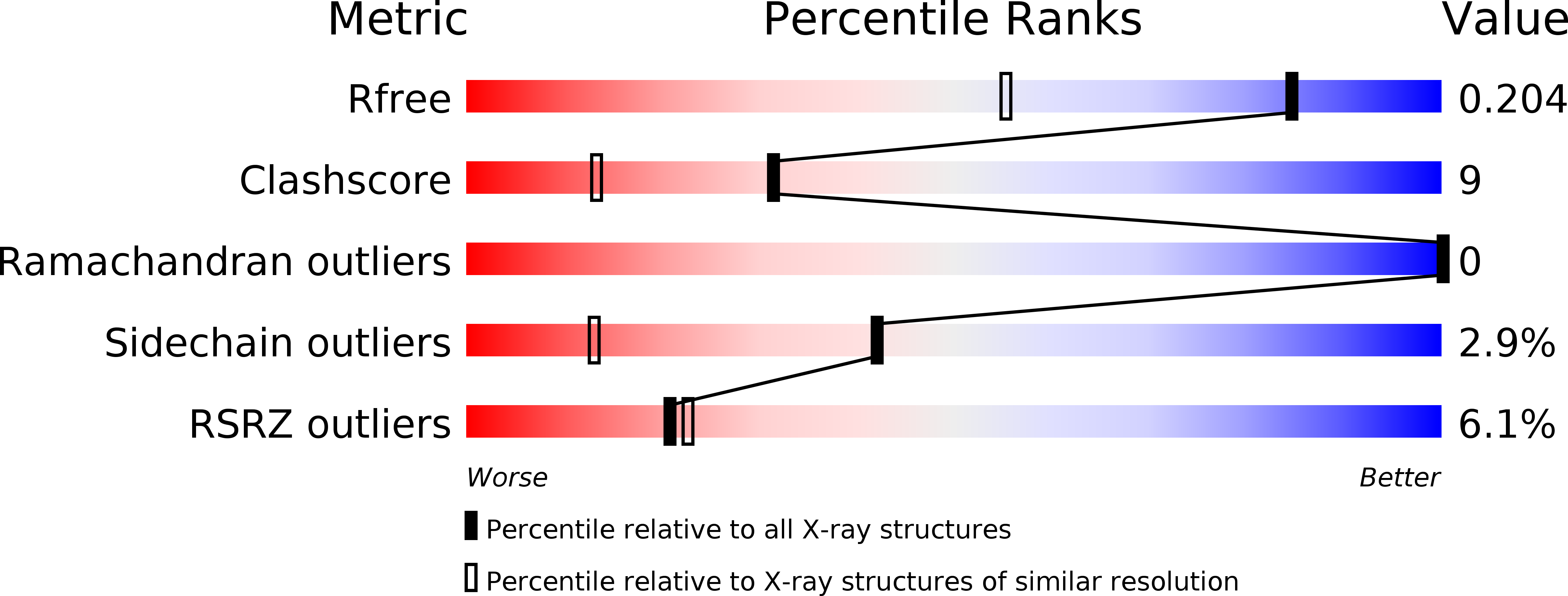
4EMT image
Entry Detail
PDB ID:
4EMT
Title:
Crystal Structure of human STING bound to c-di-GMP
Biological Source:
Source Organism:
Host Organism:
PDB Version:
Deposition Date:
2012-04-12
Release Date:
2012-06-13
Method Details:
Experimental Method:
Resolution:
1.50 Å
R-Value Free:
0.21
R-Value Work:
0.18
R-Value Observed:
0.18
Space Group:
C 2 2 21
Macromolecular Entities
Polymer Type:polypeptide(L)
Description:Transmembrane protein 173
Chain IDs:A, B
Chain Length:188
Number of Molecules:2
Biological Source:Homo sapiens
Modified Residue
Compound ID Chain ID Parent Comp ID Details 2D Image
MSE A MET SELENOMETHIONINE
Primary Citation
Structure of STING bound to cyclic di-GMP reveals the mechanism of cyclic dinucleotide recognition by the immune system.
Nat.Struct.Mol.Biol. 19 722 724 (2012)
PMID: 22728658 DOI: 10.1038/nsmb.2331

Abstact

STING (stimulator of interferon genes) is an innate immune sensor of cyclic dinucleotides that regulates the induction of type I interferons. STING's C-terminal domain forms a V-shaped dimer and binds a cyclic diguanylate monophosphate (c-di-GMP) at the dimer interface by both direct and solvent-mediated hydrogen bonds. Guanines of c-di-GMP stack against the phenolic rings of a conserved tyrosine, and mutations at the c-di-GMP binding surface reduce nucleotide binding and affect signaling.

Legend

Protein

Chemical

Disease

Primary Citation of related structures