Deposition Date
2012-04-08
Release Date
2012-07-25
Last Version Date
2024-10-16
Entry Detail
PDB ID:
4EK1
Keywords:
Title:
Crystal Structure of Electron-Spin Labeled Cytochrome P450cam
Biological Source:
Source Organism(s):
Pseudomonas putida (Taxon ID: 303)
Expression System(s):
Method Details:
Experimental Method:
Resolution:
1.97 Å
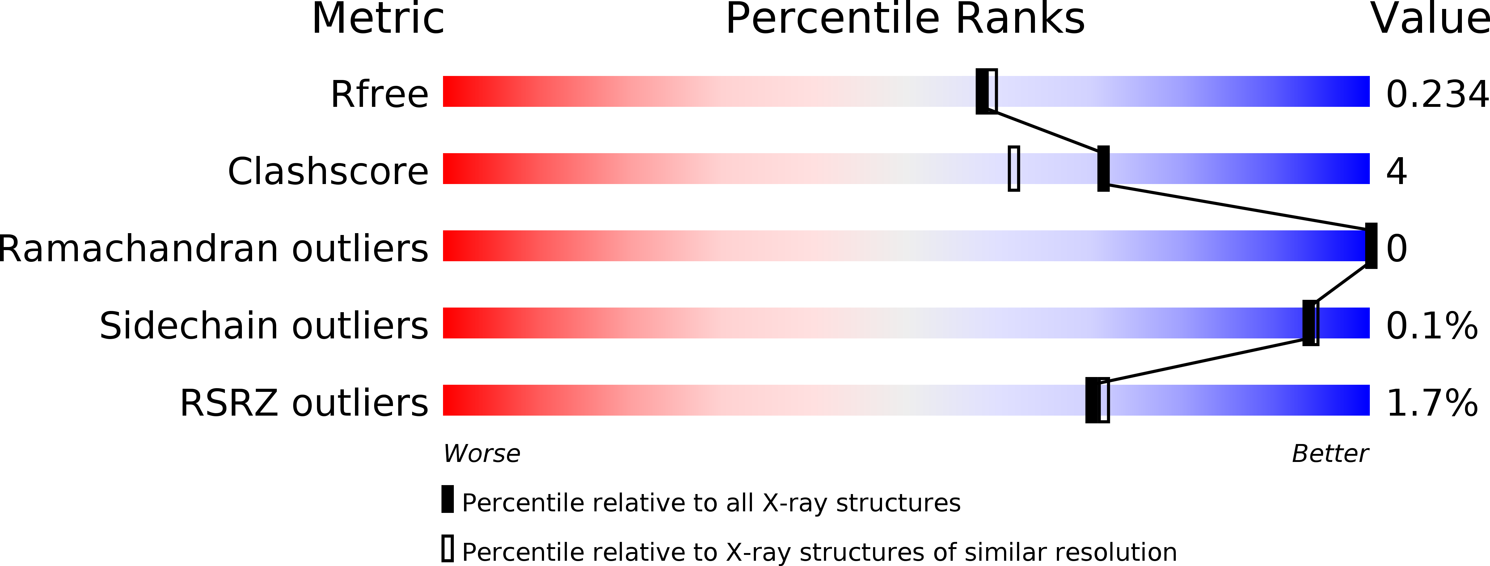
R-Value Free:
0.25
R-Value Work:
0.20
R-Value Observed:
0.20
Space Group:
P 1 21 1