Deposition Date
2012-04-06
Release Date
2013-05-01
Last Version Date
2024-02-28
Entry Detail
PDB ID:
4EJL
Keywords:
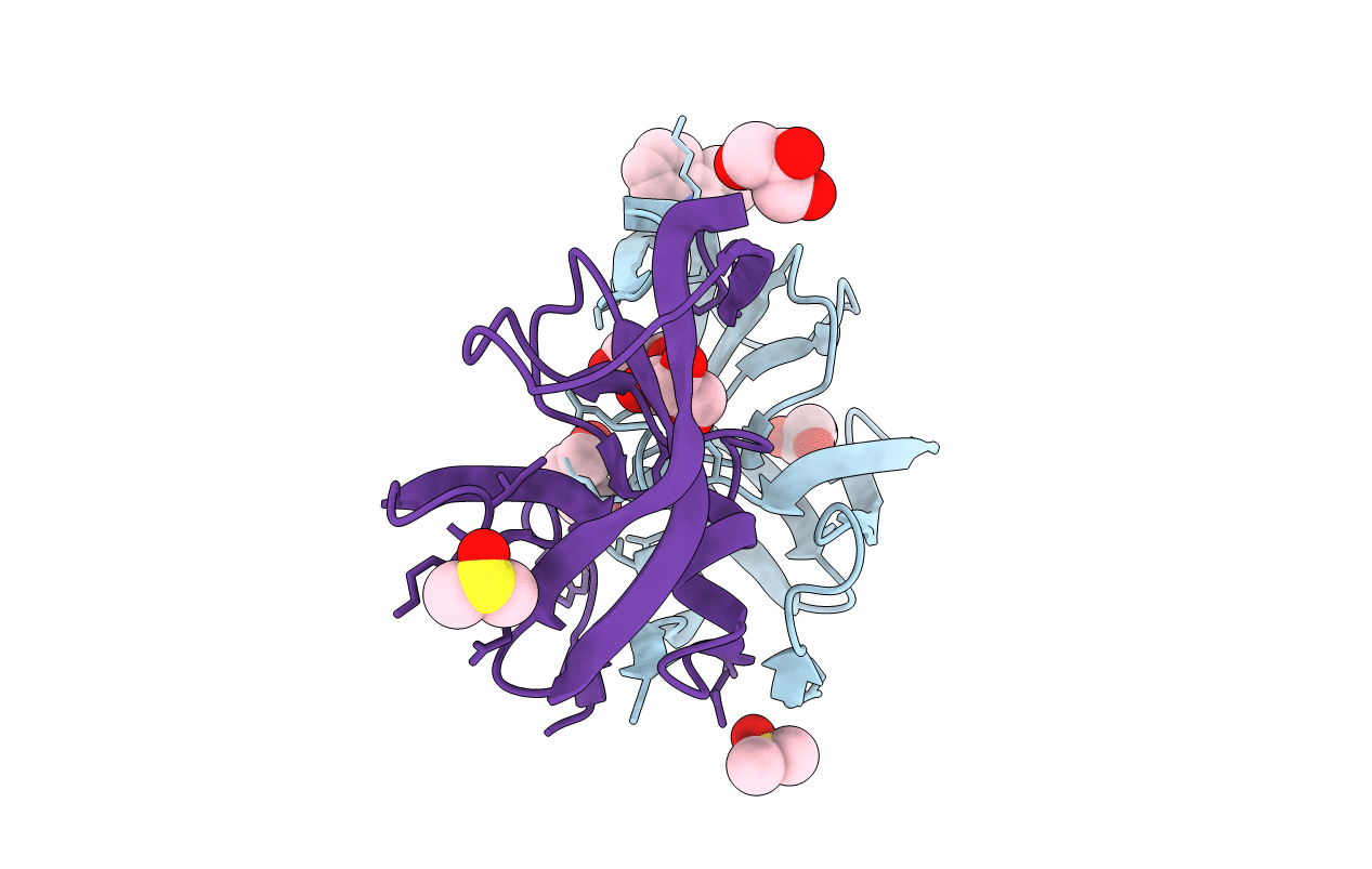
Title:
Apo HIV Protease (PR) dimer in closed form with fragment 1F1-N in the outside/top of flap
Biological Source:
Source Organism(s):
Human immunodeficiency virus 1 (Taxon ID: 11676)
Expression System(s):
Method Details:
Experimental Method:
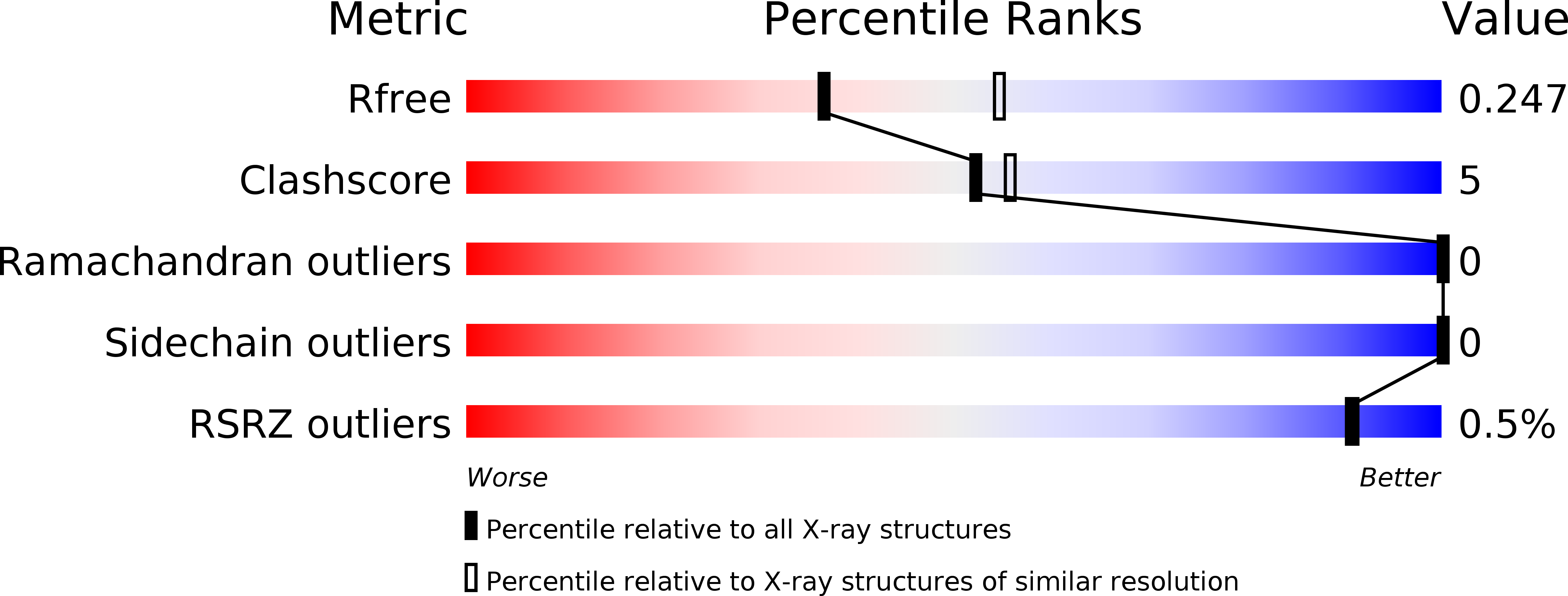
Resolution:
2.45 Å
R-Value Free:
0.24
R-Value Work:
0.19
R-Value Observed:
0.19
Space Group:
P 21 21 21