4EIJ image
Deposition Date 2012-04-05
Release Date 2013-05-08
Last Version Date 2024-02-28
Entry Detail
PDB ID:
4EIJ
Keywords:
Title:
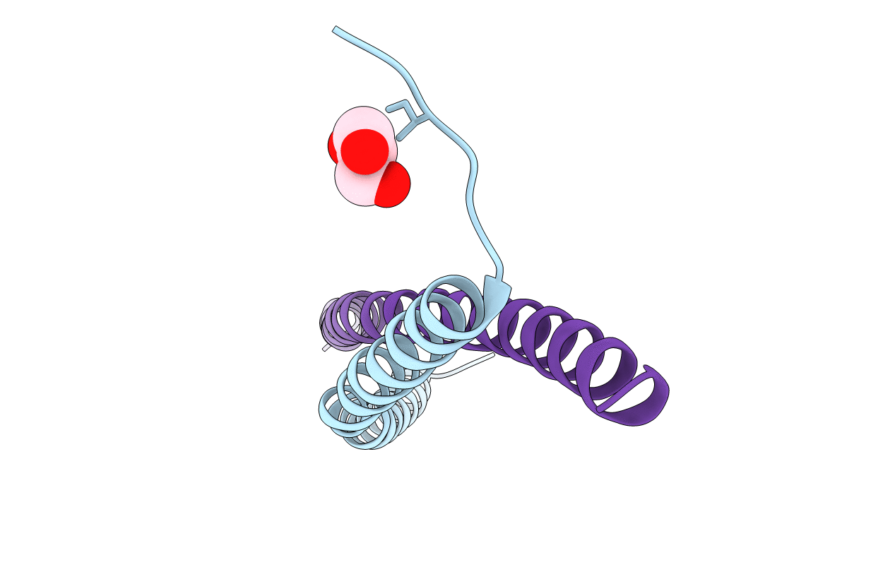
Structure of the Mumps virus phosphoprotein oligomerization domain
Biological Source:
Source Organism:
Mumps virus (Taxon ID: 11161)
Host Organism:
Method Details:
Experimental Method:
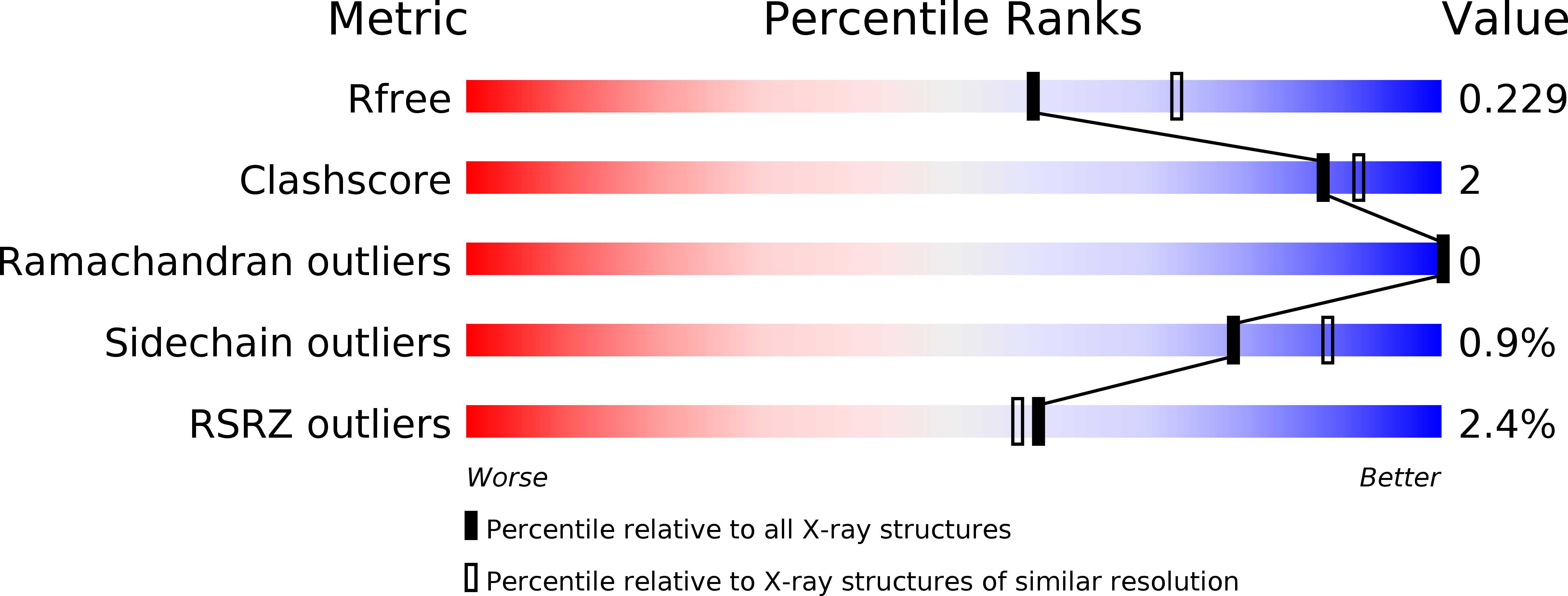
Resolution:
2.20 Å
R-Value Free:
0.23
R-Value Work:
0.18
R-Value Observed:
0.18
Space Group:
H 3 2
Macromolecular Entities
Structures with similar UniProt ID
Protein Blast
Polymer Type:polypeptide(L)
Molecule:P protein
Chain IDs:A, B
Chain Length:65
Number of Molecules:2
Biological Source:Mumps virus
Ligand Molecules
Primary Citation
Structural and functional characterization of the mumps virus phosphoprotein.
J.Virol. 87 7558 7568 (2013)
PMID: 23637399 DOI: 10.1128/JVI.00653-13

Abstact

The phosphoprotein (P) is virally encoded by the Rhabdoviridae and Paramyxoviridae in the order Mononegavirales. P is a self-associated oligomer and forms complexes with the large viral polymerase protein (L), the nucleocapsid protein (N), and the assembled nucleocapsid. P from different viruses has shown structural diversities even though their essential functions are the same. We systematically mapped the domains in mumps virus (MuV) P and investigated their interactions with nucleocapsid-like particles (NLPs). Similar to other P proteins, MuV P contains N-terminal, central, and C-terminal domains with flexible linkers between neighboring domains. By pulldown assays, we discovered that in addition to the previously proposed nucleocapsid binding domain (residues 343 to 391), the N-terminal region of MuV P (residues 1 to 194) could also bind NLPs. Further analysis of binding kinetics was conducted using surface plasmon resonance. This is the first observation that both the N- and C-terminal regions of a negative-strand RNA virus P are involved in binding the nucleocapsid. In addition, we defined the oligomerization domain (POD) of MuV P as residues 213 to 277 and determined its crystal structure. The tetrameric MuV POD is formed by one pair of long parallel α-helices with another pair in opposite orientation. Unlike the parallel orientation of each α-helix in the tetramer of Sendai virus POD, this represents a novel orientation of a POD where both the N- and the C-terminal domains are at either end of the tetramer. This is consistent with the observation that both the N- and the C-terminal domains are involved in binding the nucleocapsid.

Legend

Protein

Chemical

Disease

Primary Citation of related structures
Feedback Form
Name
Email
Institute
Feedback