4EIE image
Deposition Date 2012-04-05
Release Date 2013-04-10
Last Version Date 2025-11-12
Entry Detail
PDB ID:
4EIE
Title:
Crystal structure of cytochrome c6C from Synechococcus sp. PCC 7002
Biological Source:
Source Organism:
Host Organism:
Method Details:
Experimental Method:
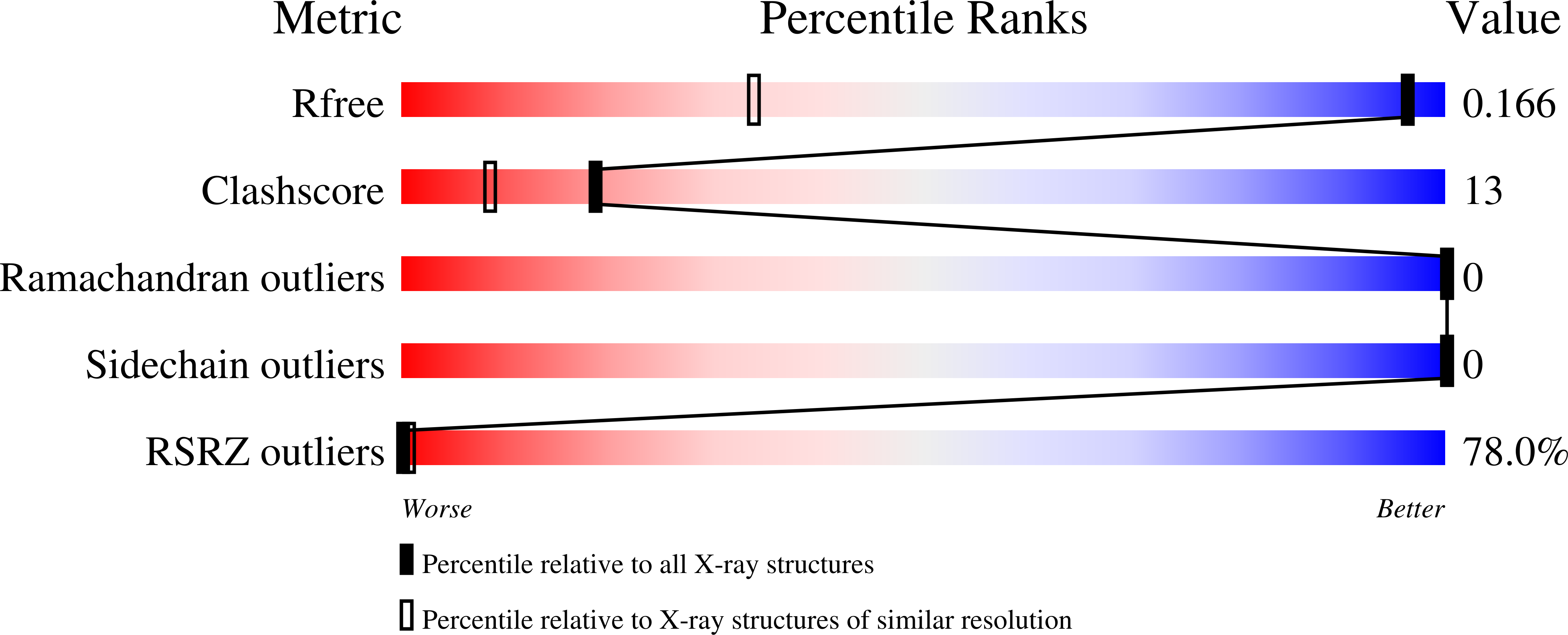
Resolution:
1.03 Å
R-Value Free:
0.16
R-Value Work:
0.13
R-Value Observed:
0.13
Space Group:
P 4 21 2
Macromolecular Entities
Structures with similar UniProt ID
Protein Blast
Polymer Type:polypeptide(L)
Molecule:Cytochrome c6
Gene (Uniprot):petJ2
Chain IDs:A
Chain Length:87
Number of Molecules:1
Biological Source:Synechococcus sp.
Primary Citation
Insights into the relationship between the haem-binding pocket and the redox potential of c6 cytochromes: four atomic resolution structures of c6 and c6-like proteins from Synechococcus sp. PCC 7002.
Acta Crystallogr.,Sect.D 70 2823 2832 (2014)
PMID: 25372674 DOI: 10.1107/S1399004714013108

Abstact

The structure of cytochrome c6C from the mesophilic cyanobacterium Synechococcus sp. PCC 7002 has been determined at 1.03 Å resolution. This is the first structural report on the recently discovered cyanobacterial cytochrome c6-like proteins found in marine and nitrogen-fixing cyanobacteria. Despite high similarity in the overall three-dimensional fold between cytochromes c6 and c6C, the latter shows saliently different electrostatic properties in terms of surface charge distribution and dipole moments. Its midpoint redox potential is less than half of the value for typical c6 cytochromes and results mainly from the substitution of one residue in the haem pocket. Here, high-resolution crystal structures of mutants of both cytochromes c6 and c6C are presented, and the impact of the mutation of specific residues in the haem-binding pocket on the redox potential is discussed. These findings contribute to the elucidation of the structure-function relationship of c6-like cytochromes.

Legend

Protein

Chemical

Disease

Primary Citation of related structures
Feedback Form
Name
Email
Institute
Feedback