4EFA image
Deposition Date 2012-03-29
Release Date 2012-10-10
Last Version Date 2023-09-13
Entry Detail
PDB ID:
4EFA
Keywords:
Title:
Crystal Structure of the Heterotrimeric EGChead Peripheral Stalk Complex of the Yeast Vacuolar ATPase - second conformation
Biological Source:
Source Organism:
Host Organism:
Method Details:
Experimental Method:
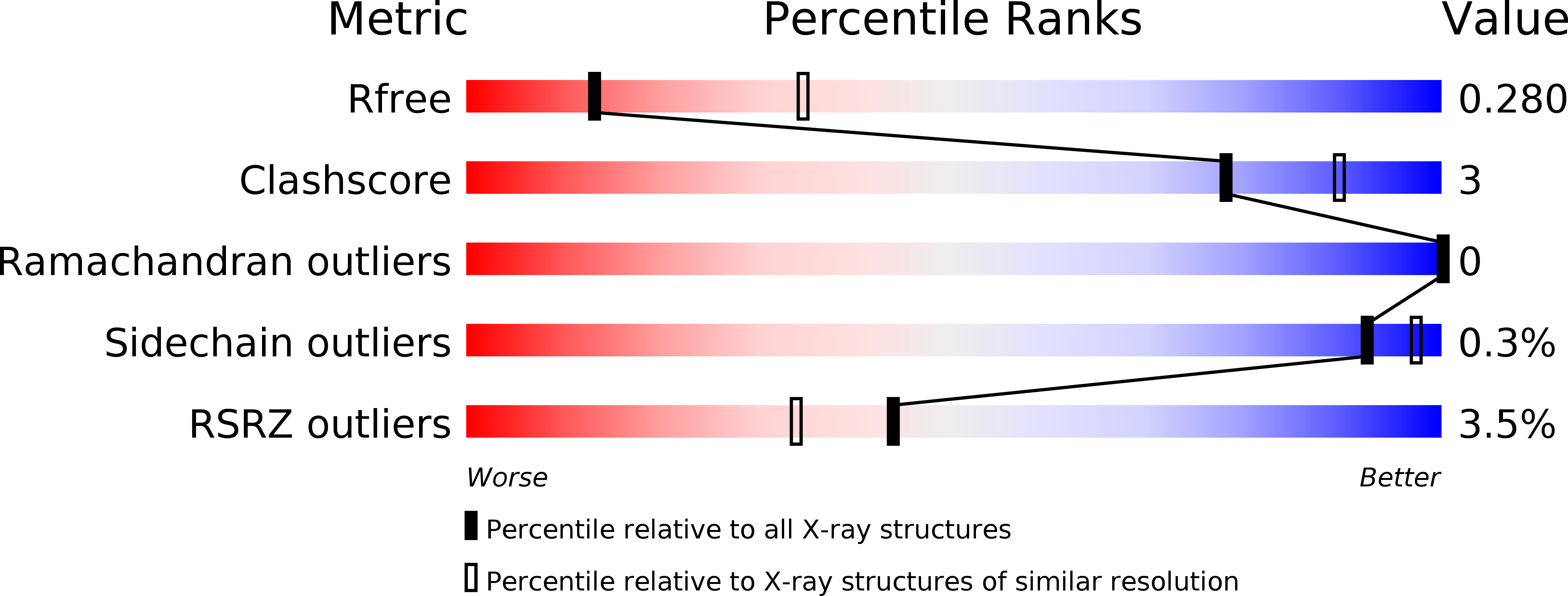
Resolution:
2.82 Å
R-Value Free:
0.27
R-Value Work:
0.22
R-Value Observed:
0.23
Space Group:
P 2 21 21
Macromolecular Entities
Structures with similar UniProt ID
Protein Blast
Polymer Type:polypeptide(L)
Molecule:V-type proton ATPase subunit C
Gene (Uniprot):VMA5
Chain IDs:A (auth: C)
Chain Length:130
Number of Molecules:1
Biological Source:Saccharomyces cerevisiae
Structures with similar UniProt ID
Protein Blast
Polymer Type:polypeptide(L)
Molecule:V-type proton ATPase subunit E
Gene (Uniprot):VMA4
Chain IDs:C (auth: E)
Chain Length:233
Number of Molecules:1
Biological Source:Saccharomyces cerevisiae
Structures with similar UniProt ID
Protein Blast
Polymer Type:polypeptide(L)
Molecule:V-type proton ATPase subunit G
Gene (Uniprot):VMA10
Chain IDs:B (auth: G)
Chain Length:119
Number of Molecules:1
Biological Source:Saccharomyces cerevisiae
Ligand Molecules
Primary Citation
Crystal Structure of the Yeast Vacuolar ATPase Heterotrimeric EGC(head) Peripheral Stalk Complex.
Structure 20 1881 1892 (2012)
PMID: 23000382 DOI: 10.1016/j.str.2012.08.020

Abstact

Vacuolar ATPases (V-ATPases) are multisubunit rotary motor proton pumps that function to acidify subcellular organelles in all eukaryotic organisms. V-ATPase is regulated by a unique mechanism that involves reversible dissociation into V₁-ATPase and V₀ proton channel, a process that involves breaking of protein interactions mediated by subunit C, the cytoplasmic domain of subunit "a" and three "peripheral stalks," each made of a heterodimer of E and G subunits. Here, we present crystal structures of a yeast V-ATPase heterotrimeric complex composed of EG heterodimer and the head domain of subunit C (C(head)). The structures show EG heterodimer folded in a noncanonical coiled coil that is stabilized at its N-terminal ends by binding to C(head). The coiled coil is disrupted by a bulge of partially unfolded secondary structure in subunit G and we speculate that this unique feature in the eukaryotic V-ATPase peripheral stalk may play an important role in enzyme structure and regulation by reversible dissociation.

Legend

Protein

Chemical

Disease

Primary Citation of related structures