4EF6 image
Deposition Date 2012-03-29
Release Date 2013-03-13
Last Version Date 2023-09-13
Entry Detail
PDB ID:
4EF6
Title:
Crystal Structure of Mycobacterium tuberculosis Pantothenate synthetase in complex with fragment 1
Biological Source:
Source Organism:
Host Organism:
Method Details:
Experimental Method:
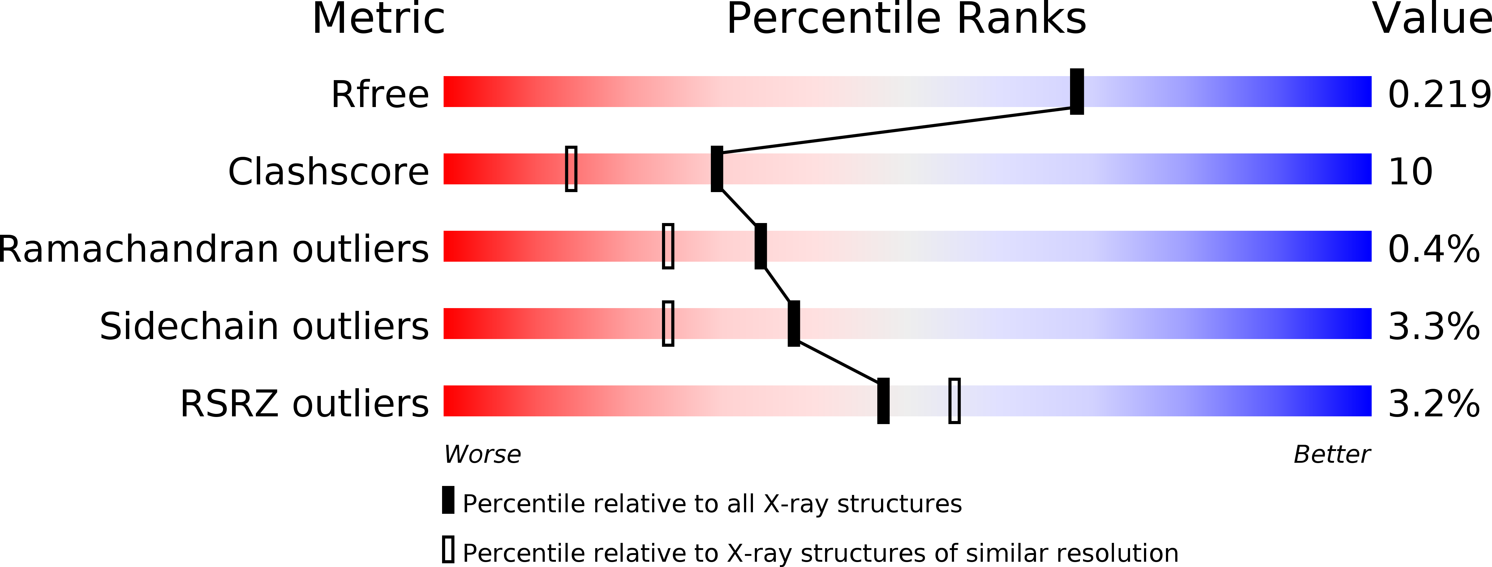
Resolution:
1.94 Å
R-Value Free:
0.21
R-Value Work:
0.16
R-Value Observed:
0.16
Space Group:
P 1 21 1
Macromolecular Entities
Polymer Type:polypeptide(L)
Molecule:Pantothenate synthetase
Mutations:T2A, G77E
Chain IDs:A, B
Chain Length:300
Number of Molecules:2
Biological Source:Mycobacterium tuberculosis
Primary Citation
Integrated biophysical approach to fragment screening and validation for fragment-based lead discovery.
Proc.Natl.Acad.Sci.USA 110 12984 12989 (2013)
PMID: 23872845 DOI: 10.1073/pnas.1304045110

Abstact

In fragment-based drug discovery, the weak affinities exhibited by fragments pose significant challenges for screening. Biophysical techniques are used to address this challenge, but there is no clear consensus on which cascade of methods is best suited to identify fragment hits that ultimately translate into bound X-ray structures and provide bona fide starting points for synthesis. We have benchmarked an integrated biophysical approach for fragment screening and validation against Mycobacterium tuberculosis pantothenate synthetase. A primary screen of 1,250 fragments library was performed by thermal shift, followed by secondary screen using one-dimensional NMR spectroscopy (water ligand observed gradient spectroscopy and saturation transfer difference binding experiments) and ultimate hit validation by isothermal titration calorimetry and X-ray crystallography. Our multibiophysical approach identified three distinct binding sites for fragments and laid a solid foundation for successful structure-based elaboration into potent inhibitors.

Legend

Protein

Chemical

Disease

Primary Citation of related structures