
Deposition Date
2012-03-28
Release Date
2012-07-04
Last Version Date
2024-10-09
Entry Detail
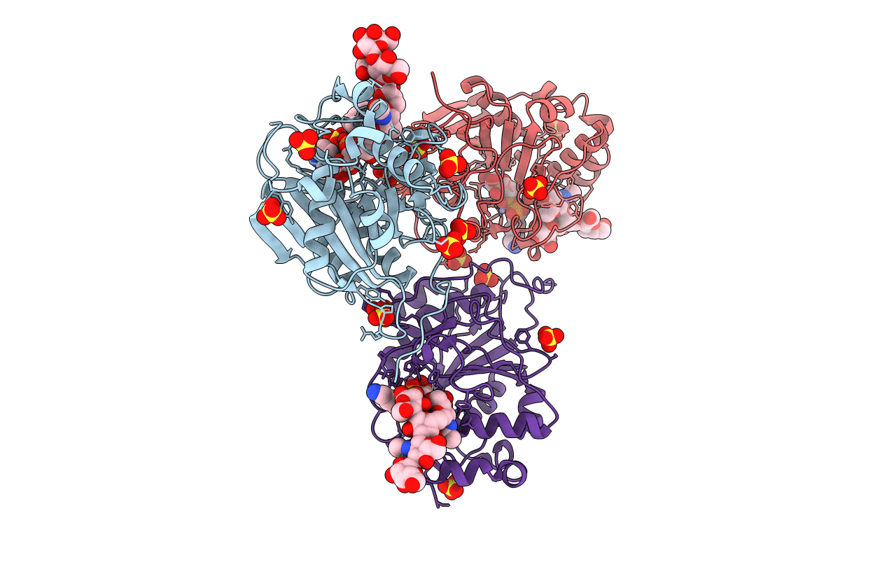
PDB ID:
4EE3
Keywords:
Title:
Crystal structure of human M340H-beta-1,4-galactosyltransferase-1 (M340H-B4GAL-T1) in complex with pentasaccharide
Biological Source:
Source Organism:
Homo sapiens (Taxon ID: 9606)
Host Organism:
Method Details:
Experimental Method:
Resolution:
2.30 Å
R-Value Free:
0.24
R-Value Work:
0.18
R-Value Observed:
0.18
Space Group:
C 2 2 21
