4EDE image
Entry Detail
PDB ID:
4EDE
Title:
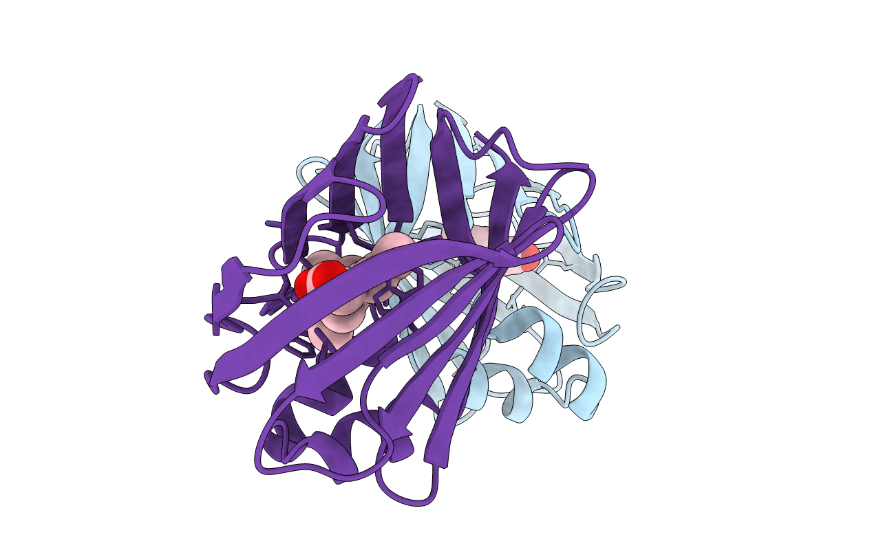
Crystal Structure of the Q108K:K40L:T51V:T53C:Y19W:R58W:T29L:A33W Mutant of Cellular Retinol Binding Protein Type II in Complex with All-trans-Retinal at 1.4 Angstrom Resolution
Biological Source:
Source Organism:
Host Organism:
PDB Version:
Deposition Date:
2012-03-27
Release Date:
2012-12-26
Method Details:
Experimental Method:
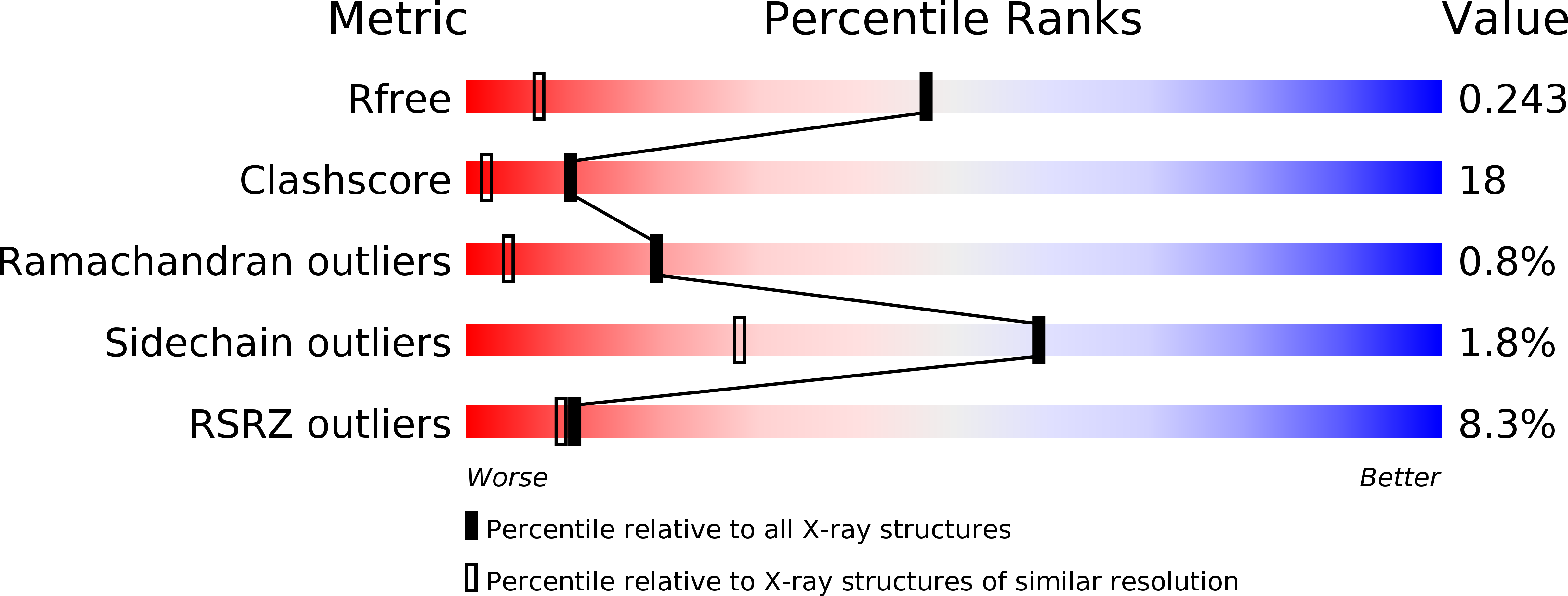
Resolution:
1.40 Å
R-Value Free:
0.24
R-Value Work:
0.19
R-Value Observed:
0.20
Space Group:
P 1
Macromolecular Entities
Polymer Type:polypeptide(L)
Description:Retinol-binding protein 2
Mutations:Q108K:K40L:T51V:T53C:Y19W:R58W:T29L:A33W
Chain IDs:A, B
Chain Length:133
Number of Molecules:2
Biological Source:Homo sapiens
Primary Citation
Tuning the electronic absorption of protein-embedded all-trans-retinal.
Science 338 1340 1343 (2012)
PMID: 23224553 DOI: 10.1126/science.1226135

Abstact

Protein-chromophore interactions are a central component of a wide variety of critical biological processes such as color vision and photosynthesis. To understand the fundamental elements that contribute to spectral tuning of a chromophore inside the protein cavity, we redesigned human cellular retinol binding protein II (hCRBPII) to fully encapsulate all-trans-retinal and form a covalent bond as a protonated Schiff base. This system, using rational mutagenesis designed to alter the electrostatic environment within the binding pocket of the host protein, enabled regulation of the absorption maximum of the pigment in the range of 425 to 644 nanometers. With only nine point mutations, the hCRBPII mutants induced a systematic shift in the absorption profile of all-trans-retinal of more than 200 nanometers across the visible spectrum.

Legend

Protein

Chemical

Disease

Primary Citation of related structures