Deposition Date
2012-03-26
Release Date
2012-07-11
Last Version Date
2024-03-20
Entry Detail
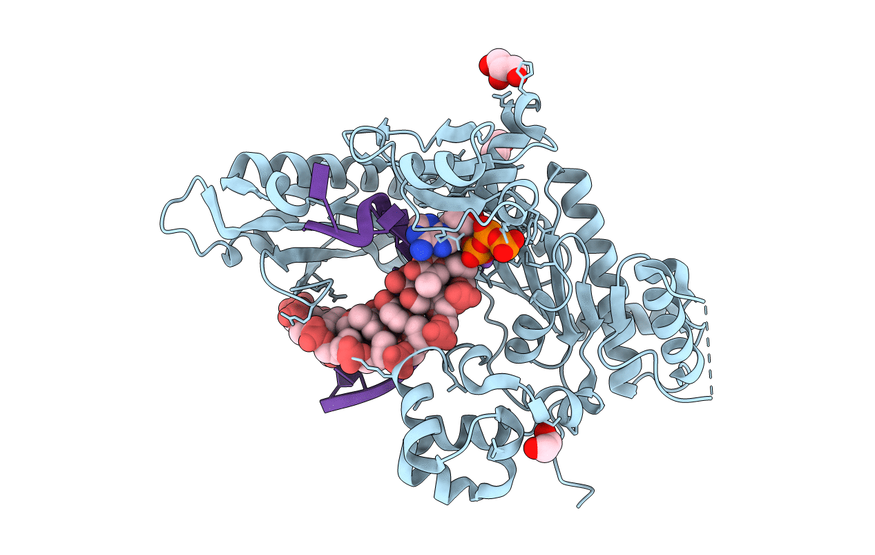
PDB ID:
4ED1
Keywords:
Title:
Human DNA polymerase eta - DNA ternary complex: AT crystal at pH 7.0 (Na+ MES) with 1 Ca2+ ion
Biological Source:
Source Organism(s):
Homo sapiens (Taxon ID: 9606)
Expression System(s):
Method Details:
Experimental Method:
Resolution:
1.81 Å
R-Value Free:
0.20
R-Value Work:
0.16
R-Value Observed:
0.17
Space Group:
P 61