4EAG image
Deposition Date 2012-03-22
Release Date 2012-06-06
Last Version Date 2023-11-08
Entry Detail
PDB ID:
4EAG
Keywords:
Title:
Co-crystal structure of an chimeric AMPK core with ATP
Biological Source:
Source Organism:
Host Organism:
Method Details:
Experimental Method:
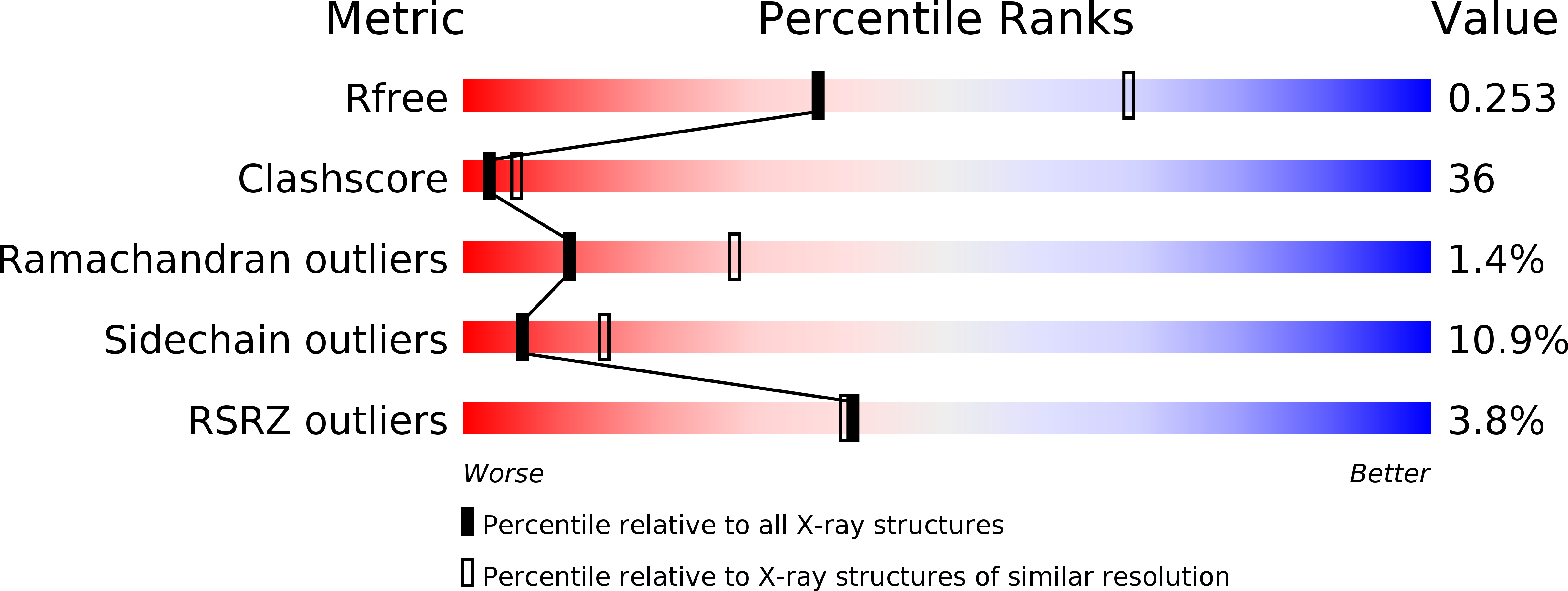
Resolution:
2.70 Å
R-Value Free:
0.25
R-Value Work:
0.20
R-Value Observed:
0.21
Space Group:
C 2 2 21
Macromolecular Entities
Structures with similar UniProt ID
Protein Blast
Polymer Type:polypeptide(L)
Molecule:EG:132E8.2 protein
Gene (Uniprot):AMPKalpha
Chain IDs:A
Chain Length:130
Number of Molecules:1
Biological Source:Drosophila melanogaster
Structures with similar UniProt ID
Protein Blast
Polymer Type:polypeptide(L)
Molecule:5'-AMP-activated protein kinase subunit beta-1
Gene (Uniprot):Prkab1
Chain IDs:B
Chain Length:85
Number of Molecules:1
Biological Source:Rattus norvegicus
Structures with similar UniProt ID
Protein Blast
Polymer Type:polypeptide(L)
Molecule:5'-AMP-activated protein kinase subunit gamma-1
Gene (Uniprot):Prkag1
Chain IDs:C
Chain Length:330
Number of Molecules:1
Biological Source:Rattus norvegicus
Primary Citation
AMP-activated protein kinase undergoes nucleotide-dependent conformational changes
Nat.Struct.Mol.Biol. 19 716 718 (2012)
PMID: 22659875 DOI: 10.1038/nsmb.2319

Abstact

The energy sensor AMP-activated protein kinase (AMPK) is a heterotrimeric complex that is allosterically activated by AMP binding to the γ subunit. Cocrystal structures of the mammalian AMPK core reveal occlusion of nucleotide-binding site 3 of the γ subunit in the presence of ATP. However, site 3 is occupied in the presence of AMP. Mutagenesis studies indicate that sites 3 and 4 are important for AMPK allosteric activation.

Legend

Protein

Chemical

Disease

Primary Citation of related structures
Feedback Form
Name
Email
Institute
Feedback