4EAB image
Deposition Date 2012-03-22
Release Date 2012-04-04
Last Version Date 2023-09-13
Entry Detail
PDB ID:
4EAB
Keywords:
Title:
X-ray crystal structure of the H141A mutant of GDP-perosamine N-acetyl transferase from Caulobacter crescentus in complex with CoA and GDP-perosamine
Biological Source:
Source Organism:
Host Organism:
Method Details:
Experimental Method:
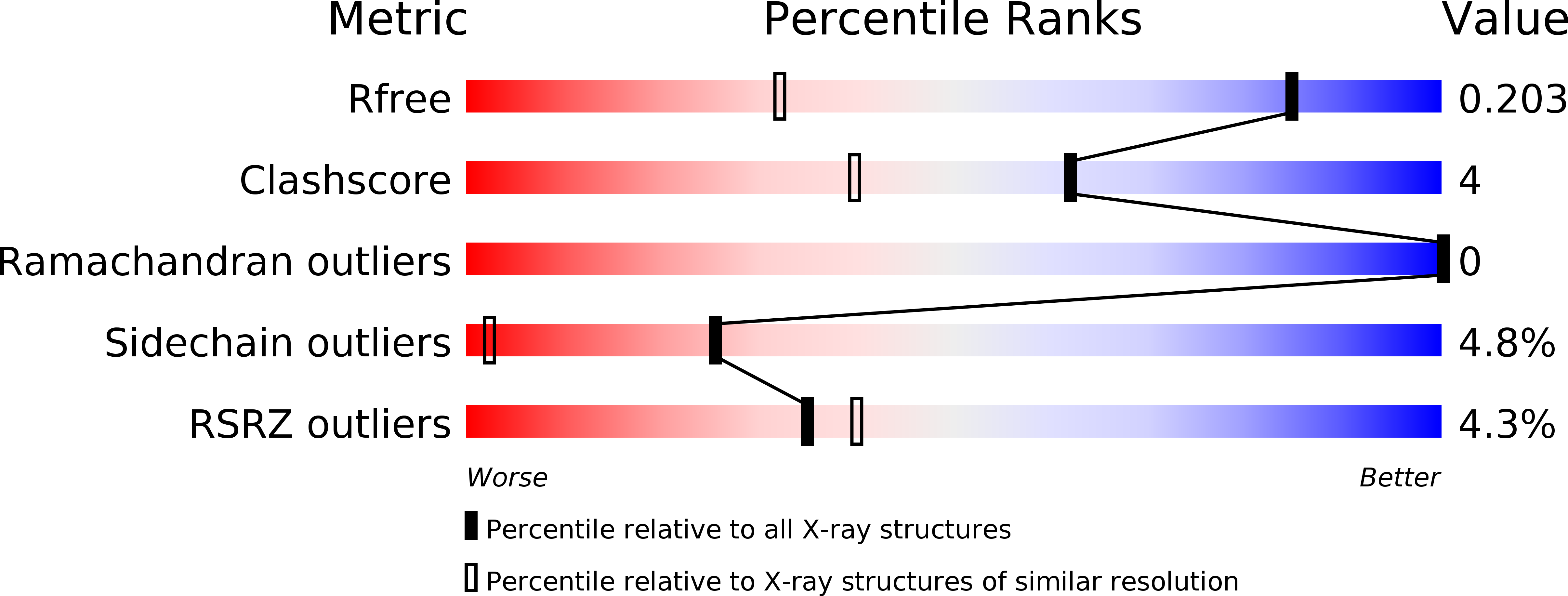
Resolution:
1.35 Å
R-Value Free:
0.20
R-Value Work:
0.18
R-Value Observed:
0.18
Space Group:
I 2 3
Macromolecular Entities
Structures with similar UniProt ID
Protein Blast
Polymer Type:polypeptide(L)
Molecule:Perosamine N-acetyltransferase
Gene (Uniprot):wbqR
Mutagens:H141A
Chain IDs:A
Chain Length:220
Number of Molecules:1
Biological Source:Caulobacter vibrioides
Primary Citation
Catalytic Mechanism of Perosamine N-Acetyltransferase Revealed by High-Resolution X-ray Crystallographic Studies and Kinetic Analyses.
Biochemistry 51 3433 3444 (2012)
PMID: 22443398 DOI: 10.1021/bi300197h

Abstact

N-Acetylperosamine is an unusual dideoxysugar found in the O-antigens of some Gram-negative bacteria, including the pathogenic Escherichia coli strain O157:H7. The last step in its biosynthesis is catalyzed by PerB, an N-acetyltransferase belonging to the left-handed β-helix superfamily of proteins. Here we describe a combined structural and functional investigation of PerB from Caulobacter crescentus. For this study, three structures were determined to 1.0 Å resolution or better: the enzyme in complex with CoA and GDP-perosamine, the protein with bound CoA and GDP-N-acetylperosamine, and the enzyme containing a tetrahedral transition state mimic bound in the active site. Each subunit of the trimeric enzyme folds into two distinct regions. The N-terminal domain is globular and dominated by a six-stranded mainly parallel β-sheet. It provides most of the interactions between the protein and GDP-perosamine. The C-terminal domain consists of a left-handed β-helix, which has nearly seven turns. This region provides the scaffold for CoA binding. On the basis of these high-resolution structures, site-directed mutant proteins were constructed to test the roles of His 141 and Asp 142 in the catalytic mechanism. Kinetic data and pH-rate profiles are indicative of His 141 serving as a general base. In addition, the backbone amide group of Gly 159 provides an oxyanion hole for stabilization of the tetrahedral transition state. The pH-rate profiles are also consistent with the GDP-linked amino sugar substrate entering the active site in its unprotonated form. Finally, for this investigation, we show that PerB can accept GDP-3-deoxyperosamine as an alternative substrate, thus representing the production of a novel trideoxysugar.

Legend

Protein

Chemical

Disease

Primary Citation of related structures
Feedback Form
Name
Email
Institute
Feedback