4E8Q image
Entry Detail
PDB ID:
4E8Q
Keywords:
Title:
Structure of Oceanobacillus iheyensis group II intron in a ligand-free state in the presence of Tl+ and Mg2+
Biological Source:
Source Organism:
PDB Version:
Deposition Date:
2012-03-20
Release Date:
2012-11-14
Method Details:
Experimental Method:
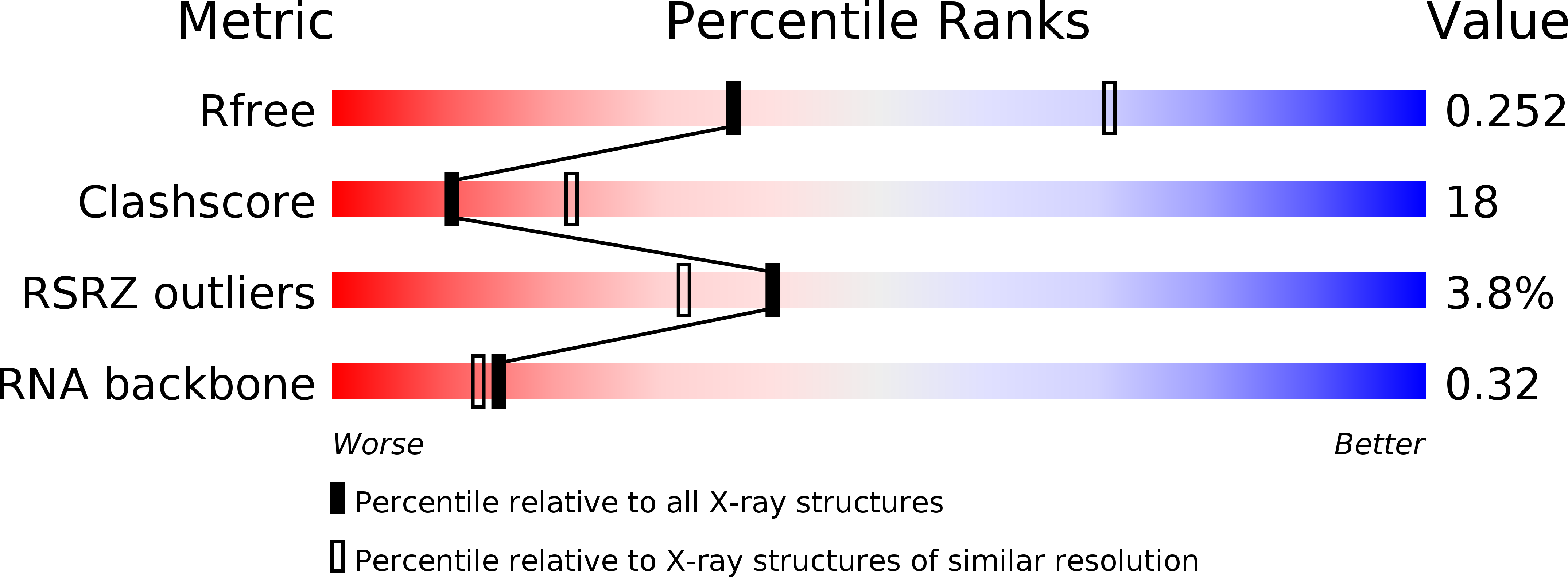
Resolution:
2.84 Å
R-Value Free:
0.25
R-Value Work:
0.20
R-Value Observed:
0.20
Space Group:
P 21 21 21
Macromolecular Entities
Polymer Type:polyribonucleotide
Description:Group IIC intron
Chain IDs:A
Chain Length:393
Number of Molecules:1
Biological Source:Oceanobacillus iheyensis
Primary Citation
Visualizing Group II Intron Catalysis through the Stages of Splicing.
Cell(Cambridge,Mass.) 151 497 507 (2012)
PMID: 23101623 DOI: 10.1016/j.cell.2012.09.033

Abstact

Group II introns are self-splicing ribozymes that share a reaction mechanism and a common ancestor with the eukaryotic spliceosome, thereby providing a model system for understanding the chemistry of pre-mRNA splicing. Here we report 14 crystal structures of a group II intron at different stages of catalysis. We provide a detailed mechanism for the first step of splicing, we describe a reversible conformational change between the first and the second steps of splicing, and we present the ligand-free intron structure after splicing in an active state that corresponds to the retrotransposable form of the intron. During each reaction, the reactants are aligned and activated by a heteronuclear four-metal-ion center that contains a metal cluster and obligate monovalent cations, and they adopt a structural arrangement similar to that of protein endonucleases. Based on our data, we propose a model for the splicing cycle and show that it is applicable to the eukaryotic spliceosome.

Legend

Protein

Chemical

Disease

Primary Citation of related structures