Deposition Date
2012-03-15
Release Date
2012-08-22
Last Version Date
2024-11-20
Entry Detail
PDB ID:
4E5Z
Keywords:
Title:
Damaged DNA induced UV-damaged DNA-binding protein (UV-DDB) dimerization and its roles in chromatinized DNA repair
Biological Source:
Source Organism(s):
Homo sapiens (Taxon ID: 9606)
Expression System(s):
Method Details:
Experimental Method:
Resolution:
3.22 Å
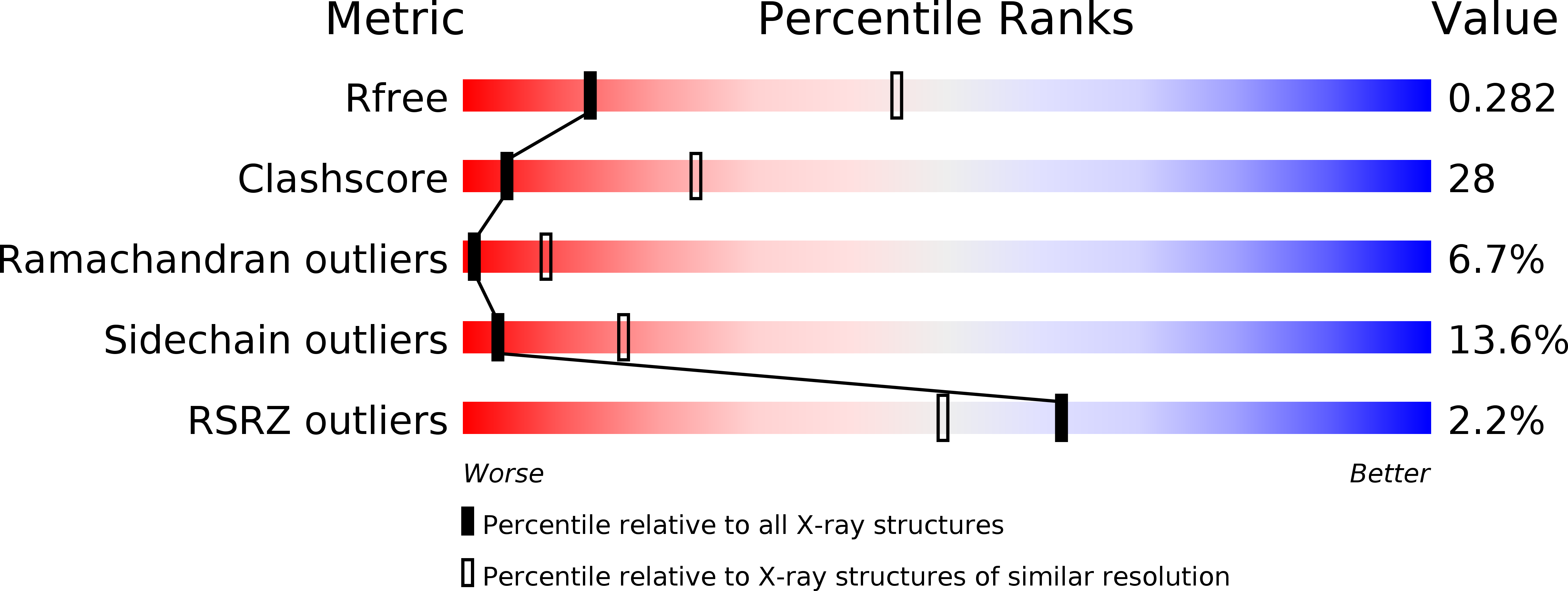
R-Value Free:
0.28
R-Value Work:
0.22
R-Value Observed:
0.23
Space Group:
P 21 2 21