Deposition Date
2012-03-12
Release Date
2012-08-22
Last Version Date
2023-09-13
Entry Detail
PDB ID:
4E4E
Keywords:
Title:
Crystal Structure of the Y34F mutant of Saccharomyces cerevisiae Manganese Superoxide Dismutase
Biological Source:
Source Organism(s):
Saccharomyces cerevisiae (Taxon ID: 4932)
Expression System(s):
Method Details:
Experimental Method:
Resolution:
1.88 Å
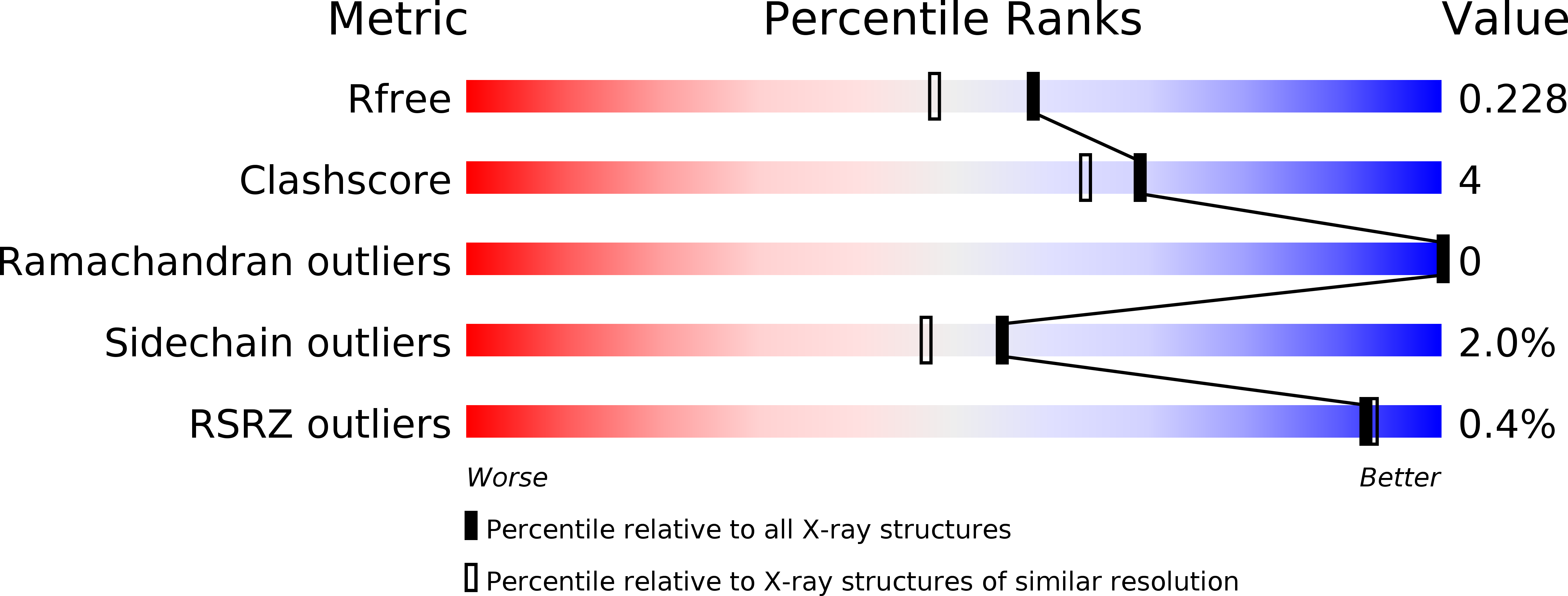
R-Value Free:
0.23
R-Value Work:
0.18
R-Value Observed:
0.19
Space Group:
P 1