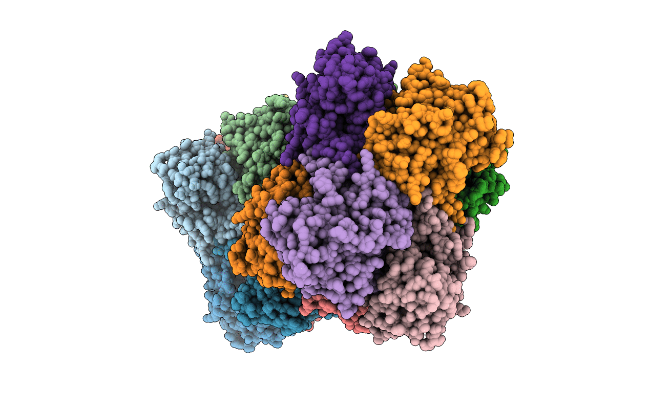
4E2Q image
Deposition Date 2012-03-09
Release Date 2012-04-18
Last Version Date 2024-03-20
Entry Detail
PDB ID:
4E2Q
Keywords:
Title:
Crystal Structure of (S)-Ureidoglycine Aminohydrolase from Arabidopsis thaliana
Biological Source:
Source Organism:
Host Organism:
Method Details:
Experimental Method:
Resolution:
2.50 Å
R-Value Free:
0.28
R-Value Work:
0.22
Space Group:
P 1 21 1
Macromolecular Entities
Polymer Type:polypeptide(L)
Molecule:Ureidoglycine aminohydrolase
Gene (Uniprot):UGLYAH
Chain IDs:A, B, C, D, E, F, G, H, I, J, K, L, M, N, O, P
Chain Length:266
Number of Molecules:16
Biological Source:Arabidopsis thaliana
Ligand Molecules
Primary Citation
Structural and functional insights into (S)-ureidoglycine aminohydrolase, key enzyme of purine catabolism in Arabidopsis thaliana
J.Biol.Chem. 287 18796 18805 (2012)
PMID: 22493446 DOI: 10.1074/jbc.M111.331819

Abstact

The ureide pathway has recently been identified as the metabolic route of purine catabolism in plants and some bacteria. In this pathway, uric acid, which is a major product of the early stage of purine catabolism, is degraded into glyoxylate and ammonia via stepwise reactions of seven different enzymes. Therefore, the pathway has a possible physiological role in mobilization of purine ring nitrogen for further assimilation. (S)-Ureidoglycine aminohydrolase enzyme converts (S)-ureidoglycine into (S)-ureidoglycolate and ammonia, providing the final substrate to the pathway. Here, we report a structural and functional analysis of this enzyme from Arabidopsis thaliana (AtUGlyAH). The crystal structure of AtUGlyAH in the ligand-free form shows a monomer structure in the bicupin fold of the β-barrel and an octameric functional unit as well as a Mn(2+) ion binding site. The structure of AtUGlyAH in complex with (S)-ureidoglycine revealed that the Mn(2+) ion acts as a molecular anchor to bind (S)-ureidoglycine, and its binding mode dictates the enantioselectivity of the reaction. Further kinetic analysis characterized the functional roles of the active site residues, including the Mn(2+) ion binding site and residues in the vicinity of (S)-ureidoglycine. These analyses provide molecular insights into the structure of the enzyme and its possible catalytic mechanism.

Legend

Protein

Chemical

Disease

Primary Citation of related structures