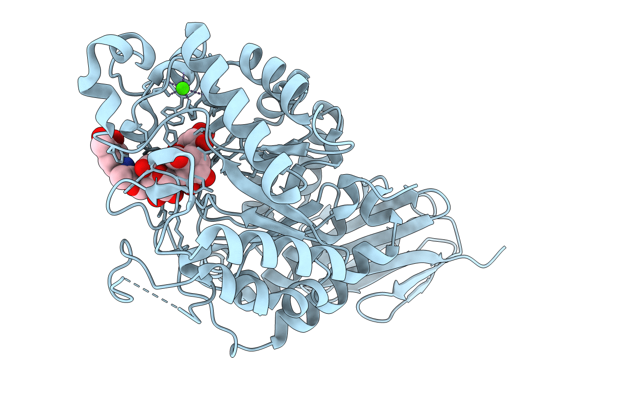
4E2O image
Entry Detail
PDB ID:
4E2O
Title:
Crystal structure of alpha-amylase from Geobacillus thermoleovorans, GTA, complexed with acarbose
Biological Source:
Host Organism:
PDB Version:
Deposition Date:
2012-03-09
Release Date:
2013-03-13
Method Details:
Experimental Method:
Resolution:
2.10 Å
R-Value Free:
0.19
R-Value Work:
0.16
R-Value Observed:
0.16
Space Group:
P 61
Macromolecular Entities
Polymer Type:polypeptide(L)
Description:Alpha-amylase
Chain IDs:A
Chain Length:454
Number of Molecules:1
Biological Source:Geobacillus thermoleovorans
Primary Citation
Crystal structure of a compact alpha-amylase from Geobacillus thermoleovorans.
Enzyme.Microb.Technol. 53 46 54 (2013)
PMID: 23683704 DOI: 10.1016/j.enzmictec.2013.03.009

Abstact

A truncated form of an α-amylase, GTA, from thermophilic Geobacillus thermoleovorans CCB_US3_UF5 was biochemically and structurally characterized. The recombinant GTA, which lacked both the N- and C-terminal transmembrane regions, functioned optimally at 70°C and pH 6.0. While enzyme activity was not enhanced by the addition of CaCl2, GTA's thermostability was significantly improved in the presence of CaCl2. The structure, in complex with an acarbose-derived pseudo-hexasaccharide, consists of the typical three domains and binds one Ca(2+) ion. This Ca(2+) ion was strongly bound and not chelated by EDTA. A predicted second Ca(2+)-binding site, however, was disordered. With limited subsites, two novel substrate-binding residues, Y147 and Y182, may help increase substrate affinity. No distinct starch-binding domain is present, although two regions rich in aromatic residues have been observed. GTA, with a smaller domain B and several shorter loops compared to other α-amylases, has one of the most compact α-amylase folds that may contribute greatly to its tight Ca(2+) binding and thermostability.

Legend

Protein

Chemical

Disease

Primary Citation of related structures