4E2H image
Deposition Date 2012-03-08
Release Date 2012-03-28
Last Version Date 2024-11-06
Entry Detail
PDB ID:
4E2H
Title:
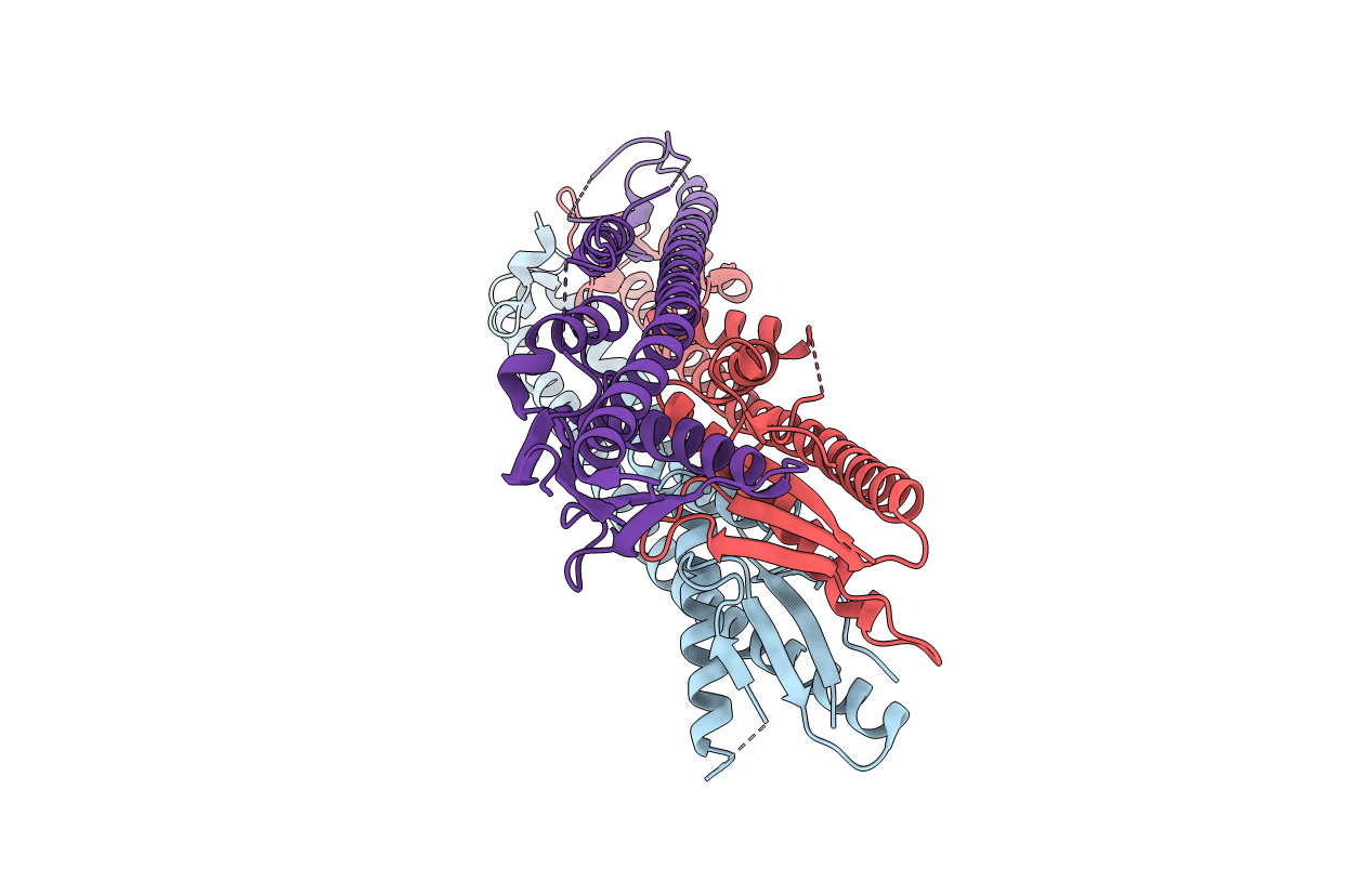
Crystal structure of the periplasmic domain of Shigella flexneri WzzB
Biological Source:
Source Organism:
Host Organism:
Method Details:
Experimental Method:
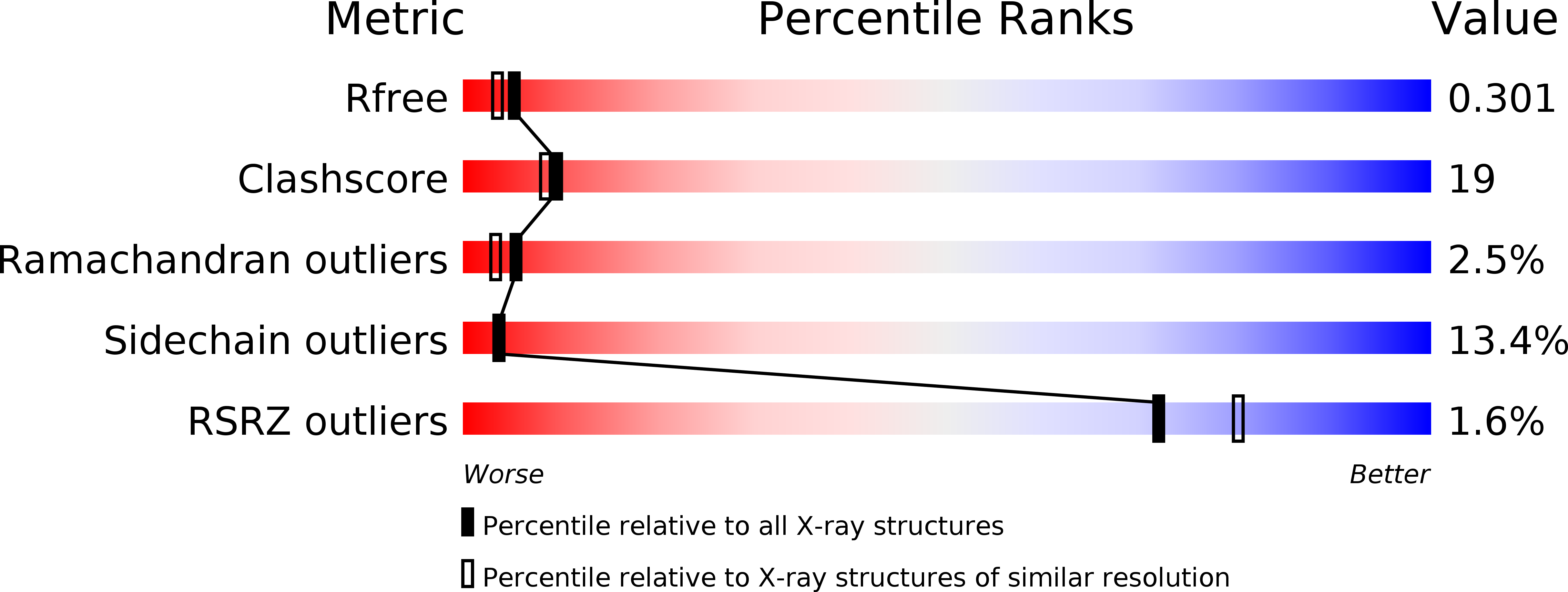
Resolution:
2.36 Å
R-Value Free:
0.30
R-Value Work:
0.24
R-Value Observed:
0.24
Space Group:
P 1 21 1
Macromolecular Entities
Polymer Type:polypeptide(L)
Molecule:Chain length determinant protein
Gene (Uniprot):wzzB
Chain IDs:A, B (auth: C), C (auth: B)
Chain Length:240
Number of Molecules:3
Biological Source:Shigella flexneri
Primary Citation
Structural Characterization of Closely Related O-antigen Lipopolysaccharide (LPS) Chain Length Regulators.
J.Biol.Chem. 287 15696 15705 (2012)
PMID: 22437828 DOI: 10.1074/jbc.M112.354837

Abstact

The surface O-antigen polymers of gram-negative bacteria exhibit a modal length distribution that depends on dedicated chain length regulator periplasmic proteins (polysaccharide co-polymerases, PCPs) anchored in the inner membrane by two transmembrane helices. In an attempt to determine whether structural changes underlie the O-antigen modal length specification, we have determined the crystal structures of several closely related PCPs, namely two chimeric PCP-1 family members solved at 1.6 and 2.8 Å and a wild-type PCP-1 from Shigella flexneri solved at 2.8 Å. The chimeric proteins form circular octamers, whereas the wild-type WzzB from S. flexneri was found to be an open trimer. We also present the structure of a Wzz(FepE) mutant, which exhibits severe attenuation in its ability to produce very long O-antigen polymers. Our findings suggest that the differences in the modal length distribution depend primarily on the surface-exposed amino acids in specific regions rather than on the differences in the oligomeric state of the PCP protomers.

Legend

Protein

Chemical

Disease

Primary Citation of related structures