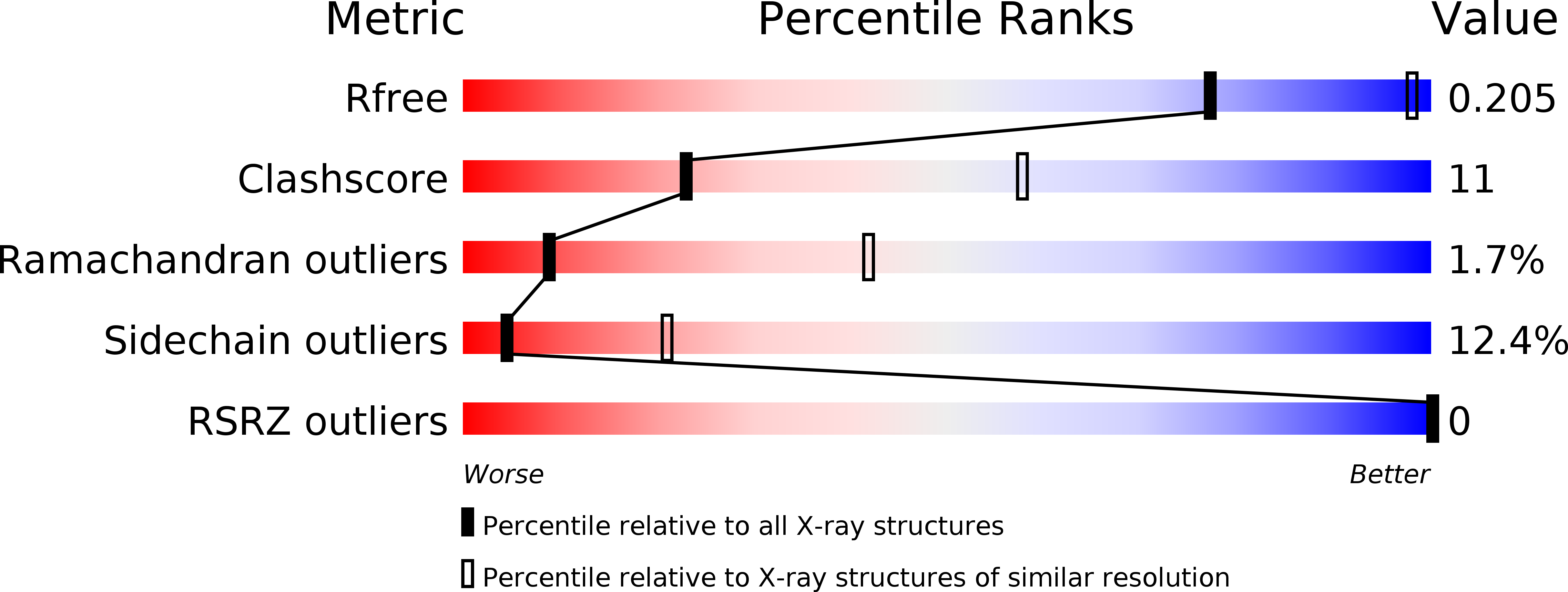
4E06 image
Deposition Date 2012-03-02
Release Date 2012-12-05
Last Version Date 2024-10-16
Entry Detail
PDB ID:
4E06
Title:
Anophelin from the malaria vector inhibits thrombin through a novel reverse-binding mechanism
Biological Source:
Source Organism:
Anopheles albimanus (Taxon ID: 7167)
Homo sapiens (Taxon ID: 9606)
Host Organism:
Method Details:
Experimental Method:
Resolution:
3.20 Å
R-Value Free:
0.20
R-Value Work:
0.17
R-Value Observed:
0.17
Space Group:
H 3 2
Macromolecular Entities
Polymer Type:polypeptide(L)
Molecule:Thrombin
Gene (Uniprot):F2
Chain IDs:B (auth: H)
Chain Length:259
Number of Molecules:1
Biological Source:Homo sapiens
Polymer Type:polypeptide(L)
Molecule:Salivary anti-thrombin peptide anophelin
Gene (Uniprot):AT
Chain IDs:C (auth: I)
Chain Length:61
Number of Molecules:1
Biological Source:Anopheles albimanus
Polymer Type:polypeptide(L)
Molecule:Thrombin
Gene (Uniprot):F2
Chain IDs:A (auth: L)
Chain Length:36
Number of Molecules:1
Biological Source:Homo sapiens
Modified Residue
Compound ID Chain ID Parent Comp ID Details 2D Image
ASN B ASN GLYCOSYLATION SITE
Primary Citation
Unique thrombin inhibition mechanism by anophelin, an anticoagulant from the malaria vector.
Proc.Natl.Acad.Sci.USA 109 E3649 E3658 (2012)
PMID: 23223529 DOI: 10.1073/pnas.1211614109

Abstact

Anopheles mosquitoes are vectors of malaria, a potentially fatal blood disease affecting half a billion humans worldwide. These blood-feeding insects include in their antihemostatic arsenal a potent thrombin inhibitor, the flexible and cysteine-less anophelin. Here, we present a thorough structure-and-function analysis of thrombin inhibition by anophelin, including the 2.3-Å crystal structure of the human thrombin·anophelin complex. Anophelin residues 32-61 are well-defined by electron density, completely occupying the long cleft between the active site and exosite I. However, in striking contrast to substrates, the D50-R53 anophelin tetrapeptide occupies the active site cleft of the enzyme, whereas the upstream residues A35-P45 shield the regulatory exosite I, defining a unique reverse-binding mode of an inhibitor to the target proteinase. The extensive interactions established, the disruption of thrombin's active site charge-relay system, and the insertion of residue R53 into the proteinase S(1) pocket in an orientation opposed to productive substrates explain anophelin's remarkable specificity and resistance to proteolysis by thrombin. Complementary biophysical and functional characterization of point mutants and truncated versions of anophelin unambiguously establish the molecular mechanism of action of this family of serine proteinase inhibitors (I77). These findings have implications for the design of novel antithrombotics.

Legend

Protein

Chemical

Disease

Primary Citation of related structures