4DWX image
Entry Detail
PDB ID:
4DWX
Keywords:
Title:
Crystal Structure of a Family GH-19 Chitinase from rye seeds
Biological Source:
Source Organism:
Host Organism:
PDB Version:
Deposition Date:
2012-02-27
Release Date:
2012-08-15
Method Details:
Experimental Method:
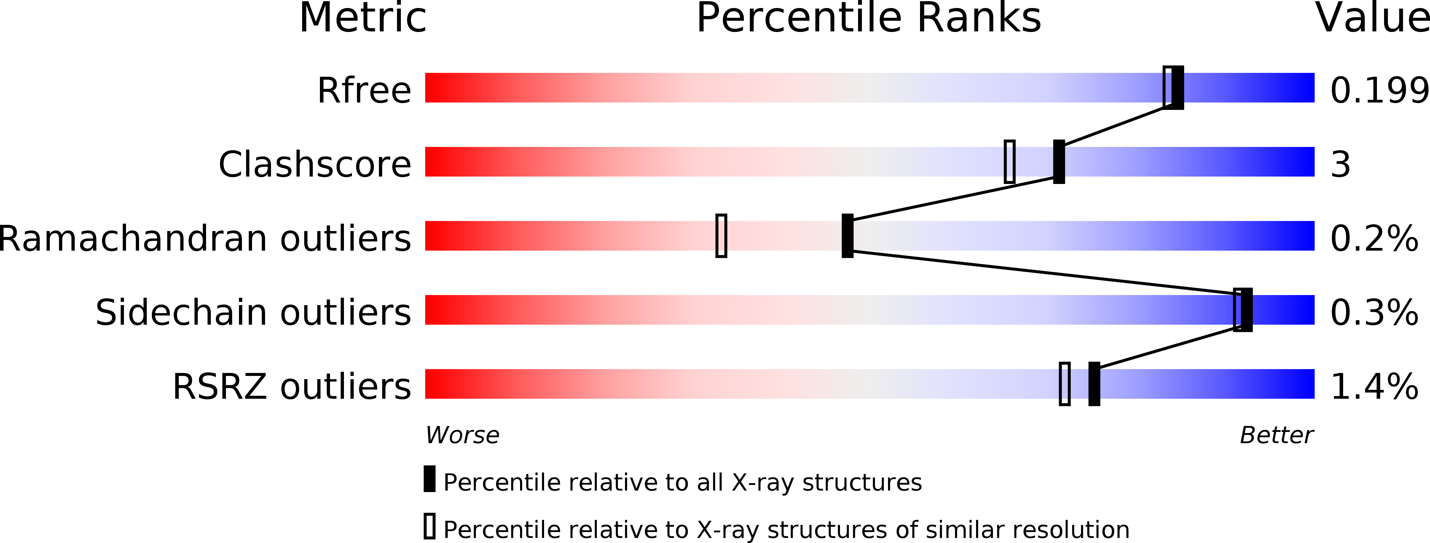
Resolution:
1.80 Å
R-Value Free:
0.20
R-Value Work:
0.17
R-Value Observed:
0.17
Space Group:
P 21 21 21
Macromolecular Entities
Polymer Type:polypeptide(L)
Description:Basic endochitinase C
Chain IDs:A, B
Chain Length:244
Number of Molecules:2
Biological Source:Secale cereale
Primary Citation
Crystal structure and chitin oligosaccharide-binding mode of a 'loopful' family GH19 chitinase from rye, Secale cereale, seeds
Febs J. 279 3639 3651 (2012)
PMID: 22831795 DOI: 10.1111/j.1742-4658.2012.08723.x

Abstact

The substrate-binding mode of a 26-kDa GH19 chitinase from rye, Secale cereale, seeds (RSC-c) was investigated by crystallography, site-directed mutagenesis and NMR spectroscopy. The crystal structure of RSC-c in a complex with an N-acetylglucosamine tetramer, (GlcNAc)(4) , was successfully solved, and revealed the binding mode of the tetramer to be an aglycon-binding site, subsites +1, +2, +3, and +4. These are the first crystallographic data showing the oligosaccharide-binding mode of a family GH19 chitinase. From HPLC analysis of the enzymatic reaction products, mutation of Trp72 to alanine was found to affect the product distribution obtained from the substrate, p-nitrophenyl penta-N-acetyl-β-chitopentaoside. Mutational experiments confirmed the crystallographic finding that the Trp72 side chain interacts with the +4 moiety of the bound substrate. To further confirm the crystallographic data, binding experiments were also conducted in solution using NMR spectroscopy. Several signals in the (1) H-(15) N HSQC spectrum of the stable isotope-labeled RSC-c were affected upon addition of (GlcNAc)(4) . Signal assignments revealed that most signals responsive to the addition of (GlcNAc)(4) are derived from amino acids located at the surface of the aglycon-binding site. The binding mode deduced from NMR binding experiments in solution was consistent with that from the crystal structure.

Legend

Protein

Chemical

Disease

Primary Citation of related structures