4DWW image
Deposition Date 2012-02-27
Release Date 2012-03-14
Last Version Date 2023-11-08
Entry Detail
PDB ID:
4DWW
Keywords:
Title:
Crystal Structure of Nattokinase from Bacillus subtilis natto
Biological Source:
Source Organism:
Method Details:
Experimental Method:
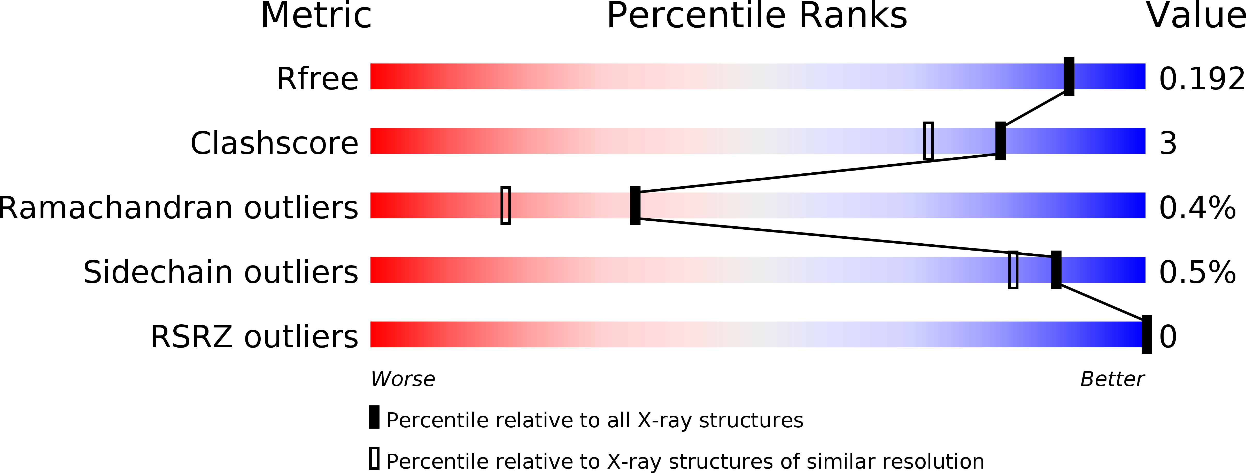
Resolution:
1.74 Å
R-Value Free:
0.19
R-Value Work:
0.13
R-Value Observed:
0.13
Space Group:
C 1 2 1
Macromolecular Entities
Polymer Type:polypeptide(L)
Molecule:Subtilisin NAT
Gene (Uniprot):aprN
Chain IDs:A
Chain Length:275
Number of Molecules:1
Biological Source:Bacillus subtilis subsp. natto
Modified Residue
Compound ID Chain ID Parent Comp ID Details 2D Image
SEB A SER O-BENZYLSULFONYL-SERINE
Ligand Molecules
Primary Citation
Purification, crystallization and preliminary X-ray diffraction experiment of nattokinase from Bacillus subtilis natto
Acta Crystallogr.,Sect.F 66 1670 1673 (2010)
PMID: 21139221 DOI: 10.1107/S1744309110043137

Abstact

Nattokinase is a single polypeptide chain composed of 275 amino acids (molecular weight 27,724) which displays strong fibrinolytic activity. Moreover, it can activate other fibrinolytic enzymes such as pro-urokinase and tissue plasminogen activator. In the present study, native nattokinase from Bacillus subtilis natto was purified using gel-filtration chromatography and crystallized to give needle-like crystals which could be used for X-ray diffraction experiments. The crystals belonged to space group C2, with unit-cell parameters a=74.3, b=49.9, c=56.3 Å, β=95.2°. Diffraction images were processed to a resolution of 1.74 Å with an Rmerge of 5.2% (15.3% in the highest resolution shell) and a completeness of 69.8% (30.0% in the highest resolution shell). This study reports the first X-ray diffraction analysis of nattokinase.

Legend

Protein

Chemical

Disease

Primary Citation of related structures