4DWS image
Deposition Date 2012-02-26
Release Date 2013-02-27
Last Version Date 2023-09-13
Entry Detail
PDB ID:
4DWS
Title:
Crystal Structure of a chitinase from the Yersinia entomophaga toxin complex
Biological Source:
Source Organism:
Host Organism:
Method Details:
Experimental Method:
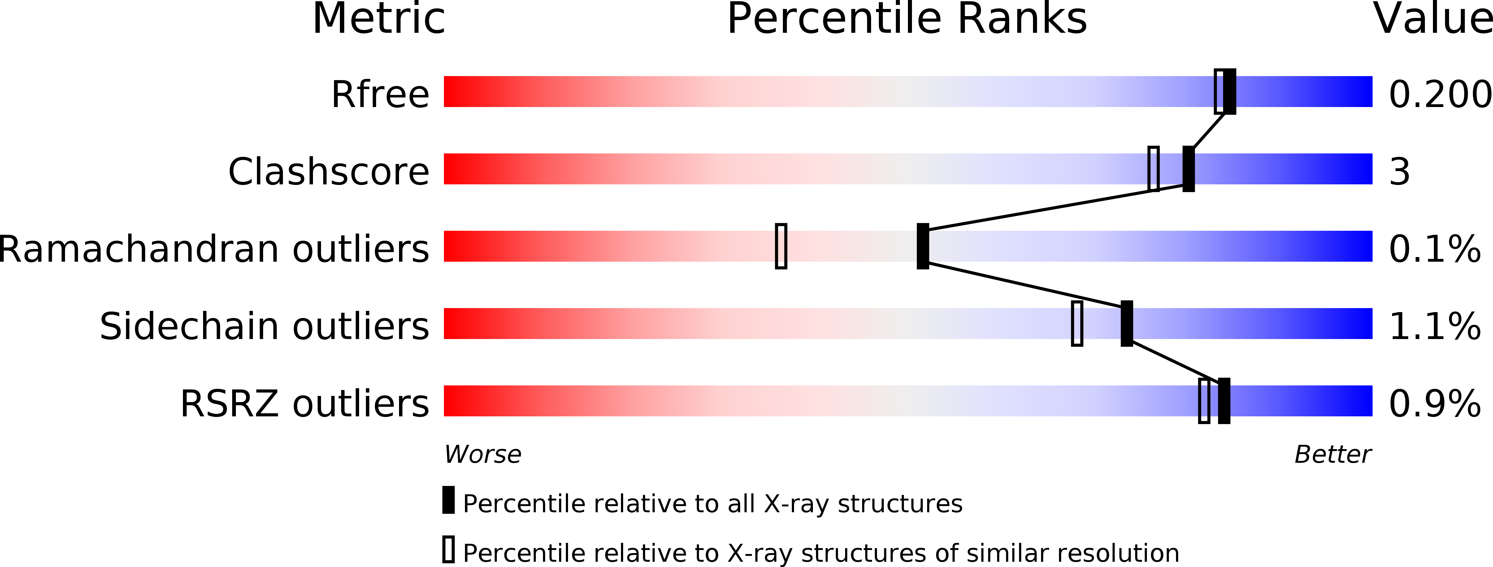
Resolution:
1.80 Å
R-Value Free:
0.19
R-Value Work:
0.15
R-Value Observed:
0.16
Space Group:
P 1 21 1
Macromolecular Entities
Polymer Type:polypeptide(L)
Molecule:Chi2
Gene (Uniprot):chi2
Chain IDs:A
Chain Length:546
Number of Molecules:1
Biological Source:Yersinia entomophaga
Polymer Type:polypeptide(L)
Molecule:Chi2
Gene (Uniprot):chi2
Chain IDs:B
Chain Length:546
Number of Molecules:1
Biological Source:Yersinia entomophaga
Polymer Type:polypeptide(L)
Molecule:Chi2
Gene (Uniprot):chi2
Chain IDs:C
Chain Length:546
Number of Molecules:1
Biological Source:Yersinia entomophaga
Polymer Type:polypeptide(L)
Molecule:Chi2
Gene (Uniprot):chi2
Chain IDs:D
Chain Length:546
Number of Molecules:1
Biological Source:Yersinia entomophaga
Modified Residue
Compound ID Chain ID Parent Comp ID Details 2D Image
M3L A LYS N-TRIMETHYLLYSINE
MLY A LYS N-DIMETHYL-LYSINE
MLZ A LYS N-METHYL-LYSINE
Ligand Molecules
Primary Citation
The BC component of ABC toxins is an RHS-repeat-containing protein encapsulation device.
Nature 501 547 550 (2013)
PMID: 23913273 DOI: 10.1038/nature12465

Abstact

The ABC toxin complexes produced by certain bacteria are of interest owing to their potent insecticidal activity and potential role in human disease. These complexes comprise at least three proteins (A, B and C), which must assemble to be fully toxic. The carboxy-terminal region of the C protein is the main cytotoxic component, and is poorly conserved between different toxin complexes. A general model of action has been proposed, in which the toxin complex binds to the cell surface via the A protein, is endocytosed, and subsequently forms a pH-triggered channel, allowing the translocation of C into the cytoplasm, where it can cause cytoskeletal disruption in both insect and mammalian cells. Toxin complexes have been visualized using single-particle electron microscopy, but no high-resolution structures of the components are available, and the role of the B protein in the mechanism of toxicity remains unknown. Here we report the three-dimensional structure of the complex formed between the B and C proteins, determined to 2.5 Å by X-ray crystallography. These proteins assemble to form an unprecedented, large hollow structure that encapsulates and sequesters the cytotoxic, C-terminal region of the C protein like the shell of an egg. The shell is decorated on one end by a β-propeller domain, which mediates attachment of the B-C heterodimer to the A protein in the native complex. The structure reveals how C auto-proteolyses when folded in complex with B. The C protein is the first example, to our knowledge, of a structure that contains rearrangement hotspot (RHS) repeats, and illustrates a marked structural architecture that is probably conserved across both this widely distributed bacterial protein family and the related eukaryotic tyrosine-aspartate (YD)-repeat-containing protein family, which includes the teneurins. The structure provides the first clues about the function of these protein repeat families, and suggests a generic mechanism for protein encapsulation and delivery.

Legend

Protein

Chemical

Disease

Primary Citation of related structures