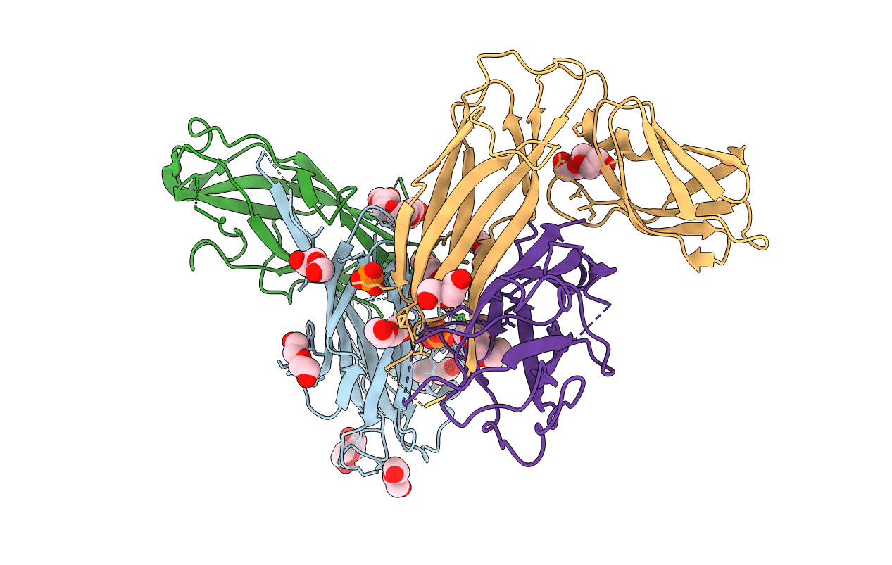
4DWH image
Entry Detail
PDB ID:
4DWH
Title:
Structure of the major type 1 pilus subunit FIMA bound to the FIMC (2.5 A resolution)
Biological Source:
Source Organism:
Host Organism:
PDB Version:
Deposition Date:
2012-02-24
Release Date:
2012-05-30
Method Details:
Experimental Method:
Resolution:
2.50 Å
R-Value Free:
0.29
R-Value Work:
0.23
R-Value Observed:
0.23
Space Group:
C 1 2 1
Macromolecular Entities
Polymer Type:polypeptide(L)
Description:Chaperone protein fimC
Chain IDs:A, C
Chain Length:205
Number of Molecules:2
Biological Source:Escherichia coli
Polymer Type:polypeptide(L)
Description:Type-1 fimbrial protein, A chain
Chain IDs:B, D
Chain Length:143
Number of Molecules:2
Biological Source:Escherichia coli
Primary Citation
Quality control of disulfide bond formation in pilus subunits by the chaperone FimC.
Nat.Chem.Biol. 8 707 713 (2012)
PMID: 22772153 DOI: 10.1038/nchembio.1019

Abstact

Type 1 pili from uropathogenic Escherichia coli are filamentous, noncovalent protein complexes mediating bacterial adhesion to the host tissue. All structural pilus subunits are homologous proteins sharing an invariant disulfide bridge. Here we show that disulfide bond formation in the unfolded subunits, catalyzed by the periplasmic oxidoreductase DsbA, is required for subunit recognition by the assembly chaperone FimC and for FimC-catalyzed subunit folding. FimC thus guarantees quantitative disulfide bond formation in each of the up to 3,000 subunits of the pilus. The X-ray structure of the complex between FimC and the main pilus subunit FimA and the kinetics of FimC-catalyzed FimA folding indicate that FimC accelerates folding of pilus subunits by lowering their topological complexity. The kinetic data, together with the measured in vivo concentrations of DsbA and FimC, predict an in vivo half-life of 2 s for oxidative folding of FimA in the periplasm.

Legend

Protein

Chemical

Disease

Primary Citation of related structures