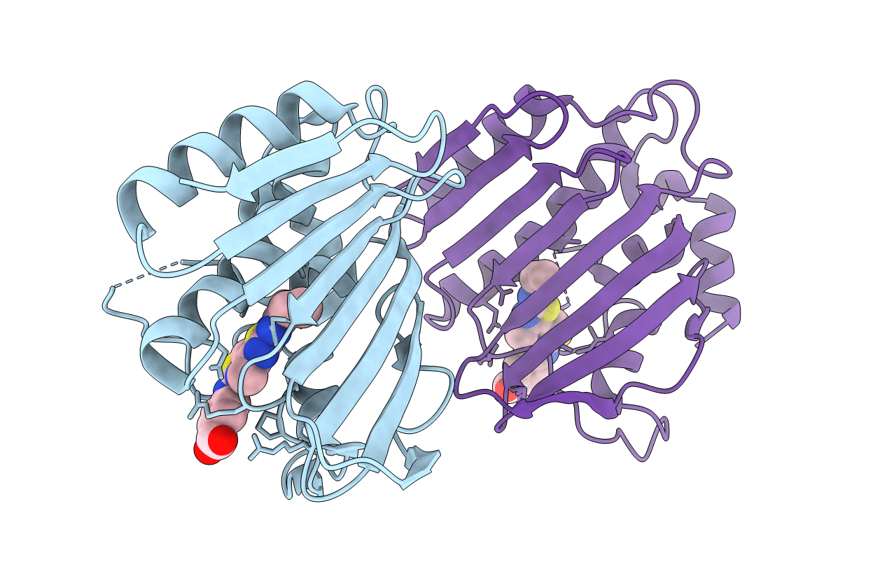
4DUH image
Entry Detail
PDB ID:
4DUH
Title:
Crystal structure of 24 kDa domain of E. coli DNA gyrase B in complex with small molecule inhibitor
Biological Source:
Source Organism:
Host Organism:
PDB Version:
Deposition Date:
2012-02-22
Release Date:
2012-08-01
Method Details:
Experimental Method:
Resolution:
1.50 Å
R-Value Free:
0.22
R-Value Work:
0.19
R-Value Observed:
0.19
Space Group:
P 1 21 1
Macromolecular Entities
Polymer Type:polypeptide(L)
Description:DNA gyrase subunit B
Chain IDs:A, B
Chain Length:220
Number of Molecules:2
Biological Source:Escherichia coli
Ligand Molecules
Primary Citation
Structure-based discovery of substituted 4,5'-bithiazoles as novel DNA gyrase inhibitors.
J.Med.Chem. 55 6413 6426 (2012)
PMID: 22731783 DOI: 10.1021/jm300395d

Abstact

Bacterial DNA gyrase is a well-established and validated target for the development of novel antibacterials. Starting from the available structural information about the binding of the natural product inhibitor, clorobiocin, we identified a novel series of 4'-methyl-N(2)-phenyl-[4,5'-bithiazole]-2,2'-diamine inhibitors of gyrase B with a low micromolar inhibitory activity by implementing a two-step structure-based design procedure. This novel class of DNA gyrase inhibitors was extensively investigated by various techniques (differential scanning fluorimetry, surface plasmon resonance, and microscale thermophoresis). The binding mode of the potent inhibitor 18 was revealed by X-ray crystallography, confirming our initial in silico binding model. Furthermore, the high resolution of the complex structure allowed for the placement of the Gly97-Ser108 flexible loop, thus revealing its role in binding of this class of compounds. The crystal structure of the complex protein G24 and inhibitor 18 provides valuable information for further optimization of this novel class of DNA gyrase B inhibitors.

Legend

Protein

Chemical

Disease

Primary Citation of related structures