4DTZ image
Entry Detail
PDB ID:
4DTZ
Keywords:
Title:
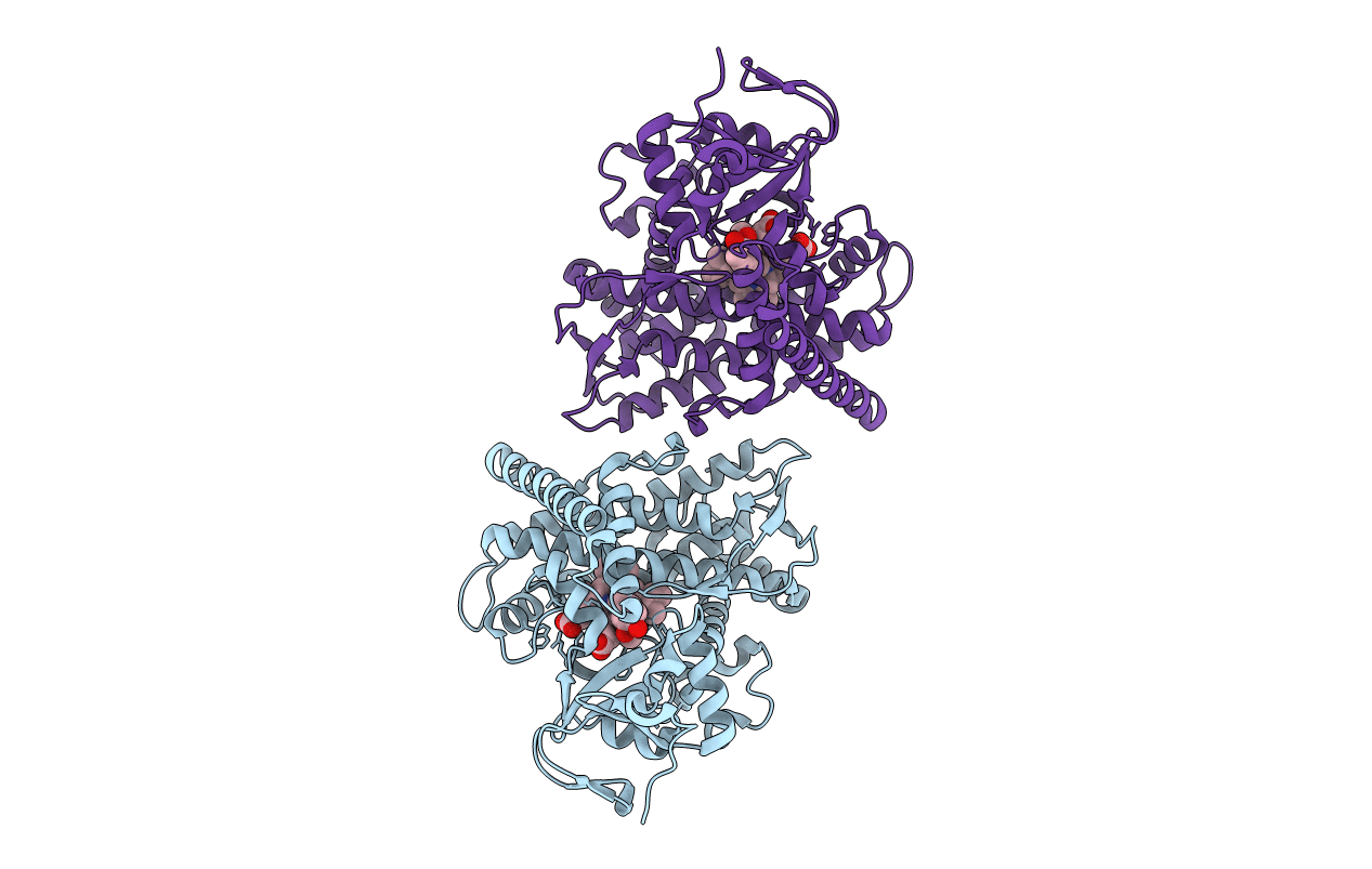
cytochrome P450 BM3h-8C8 MRI sensor bound to dopamine
Biological Source:
Source Organism:
Host Organism:
PDB Version:
Deposition Date:
2012-02-21
Release Date:
2012-06-13
Method Details:
Experimental Method:
Resolution:
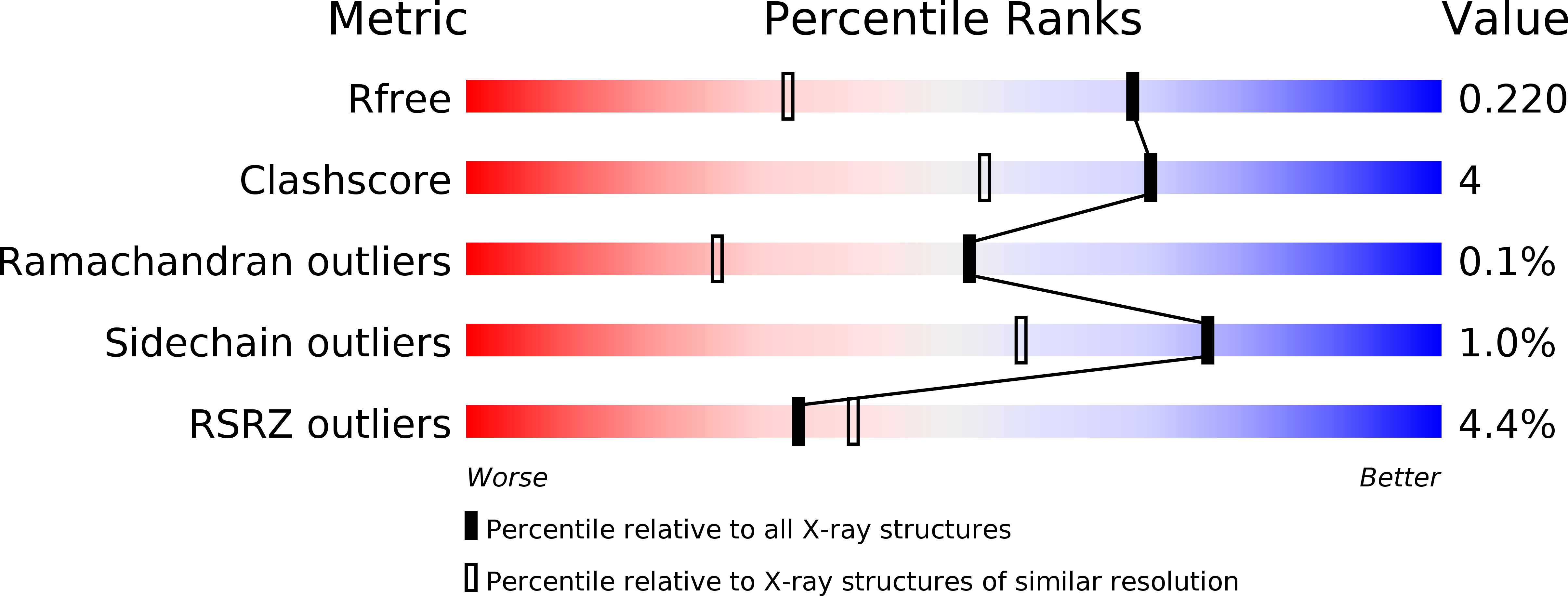
1.55 Å
R-Value Free:
0.22
R-Value Work:
0.17
R-Value Observed:
0.17
Space Group:
P 1 21 1
Macromolecular Entities
Polymer Type:polypeptide(L)
Description:cytochrome P450 BM3 variant 8C8
Mutations:L75P, Q189R, I263A, T268A, V286E
Chain IDs:A, B
Chain Length:469
Number of Molecules:2
Biological Source:Bacillus megaterium
Primary Citation
Structure-guided directed evolution of highly selective p450-based magnetic resonance imaging sensors for dopamine and serotonin.
J.Mol.Biol. 422 245 262 (2012)
PMID: 22659321 DOI: 10.1016/j.jmb.2012.05.029

Abstact

New tools that allow dynamic visualization of molecular neural events are important for studying the basis of brain activity and disease. Sensors that permit ligand-sensitive magnetic resonance imaging (MRI) are useful reagents due to the noninvasive nature and good temporal and spatial resolution of MR methods. Paramagnetic metalloproteins can be effective MRI sensors due to the selectivity imparted by the protein active site and the ability to tune protein properties using techniques such as directed evolution. Here, we show that structure-guided directed evolution of the active site of the cytochrome P450-BM3 heme domain produces highly selective MRI probes with submicromolar affinities for small molecules. We report a new, high-affinity dopamine sensor as well as the first MRI reporter for serotonin, with which we demonstrate quantification of neurotransmitter release in vitro. We also present a detailed structural analysis of evolved cytochrome P450-BM3 heme domain lineages to systematically dissect the molecular basis of neurotransmitter binding affinity, selectivity, and enhanced MRI contrast activity in these engineered proteins.

Legend

Protein

Chemical

Disease

Primary Citation of related structures