4DT7 image
Deposition Date 2012-02-20
Release Date 2012-05-09
Last Version Date 2024-11-06
Entry Detail
PDB ID:
4DT7
Keywords:
Title:
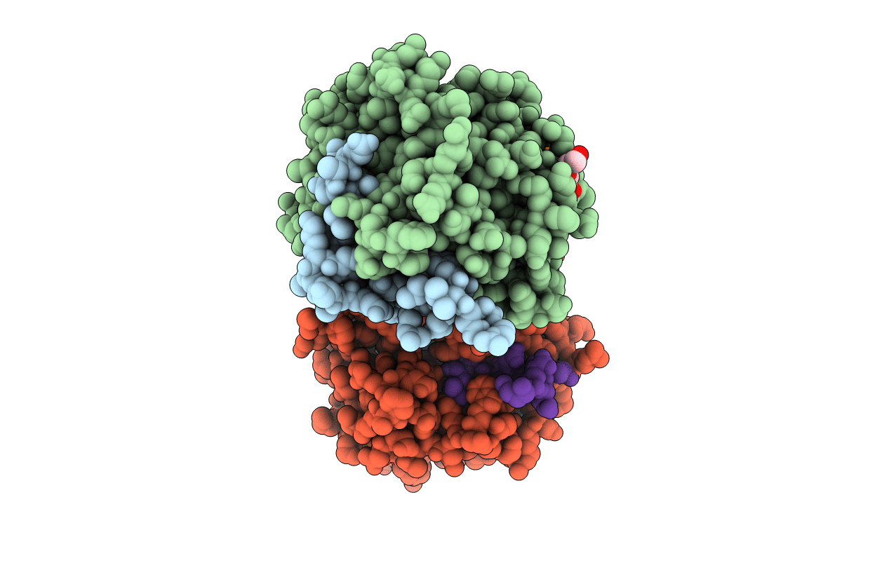
Crystal structure of thrombin bound to the activation domain QEDQVDPRLIDGKMTRRGDS of protein C
Biological Source:
Source Organism:
Homo sapiens (Taxon ID: 9606)
Host Organism:
Method Details:
Experimental Method:
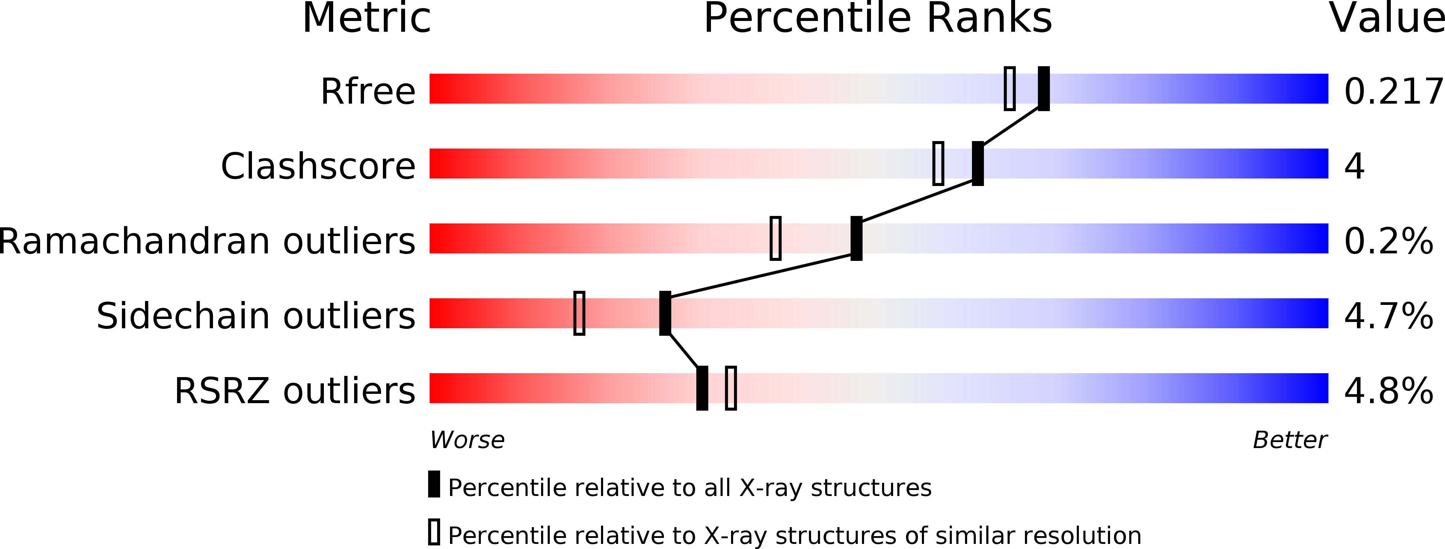
Resolution:
1.90 Å
R-Value Free:
0.21
R-Value Work:
0.17
R-Value Observed:
0.17
Space Group:
P 1 21 1
Macromolecular Entities
Structures with similar UniProt ID
Protein Blast
Polymer Type:polypeptide(L)
Molecule:Thrombin light chain
Gene (Uniprot):F2
Chain IDs:A, C
Chain Length:32
Number of Molecules:2
Biological Source:Homo sapiens
Structures with similar UniProt ID
Protein Blast
Polymer Type:polypeptide(L)
Molecule:Thrombin heavy chain
Gene (Uniprot):F2
Mutagens:S195A
Chain IDs:B, D
Chain Length:259
Number of Molecules:2
Biological Source:Homo sapiens
Structures with similar UniProt ID
Protein Blast
Polymer Type:polypeptide(L)
Molecule:Vitamin K-dependent protein C
Gene (Uniprot):PROC
Chain IDs:E, F
Chain Length:20
Number of Molecules:2
Biological Source:Homo sapiens
Primary Citation
Exposure of R169 controls protein C activation and autoactivation.
Blood 120 664 670 (2012)
PMID: 22535660 DOI: 10.1182/blood-2012-03-415323

Abstact

Protein C is activated by thrombin with a value of k(cat)/K(m) = 0.11mM(-1)s(-1) that increases 1700-fold in the presence of the cofactor thrombomodulin. The molecular origin of this effect triggering an important feedback loop in the coagulation cascade remains elusive. Acidic residues in the activation domain of protein C are thought to electrostatically clash with the active site of thrombin. However, functional and structural data reported here support an alternative scenario. The thrombin precursor prethrombin-2 has R15 at the site of activation in ionic interaction with E14e, D14l, and E18, instead of being exposed to solvent for proteolytic attack. Residues E160, D167, and D172 around the site of activation at R169 of protein C occupy the same positions as E14e, D14l, and E18 in prethrombin-2. Caging of R169 by E160, D167, and D172 is responsible for much of the poor activity of thrombin toward protein C. The E160A/D167A/D172A mutant is activated by thrombin 63-fold faster than wild-type in the absence of thrombomodulin and, over a slower time scale, spontaneously converts to activated protein C. These findings establish a new paradigm for cofactor-assisted reactions in the coagulation cascade.

Legend

Protein

Chemical

Disease

Primary Citation of related structures
Feedback Form
Name
Email
Institute
Feedback