4DR5 image
Entry Detail
PDB ID:
4DR5
Title:
Crystal structure of the Thermus thermophilus (HB8) 30S ribosomal subunit with codon, crystallographically disordered cognate transfer RNA anticodon stem-loop and streptomycin bound
Biological Source:
Source Organism:
PDB Version:
Deposition Date:
2012-02-16
Release Date:
2012-11-14
Method Details:
Experimental Method:
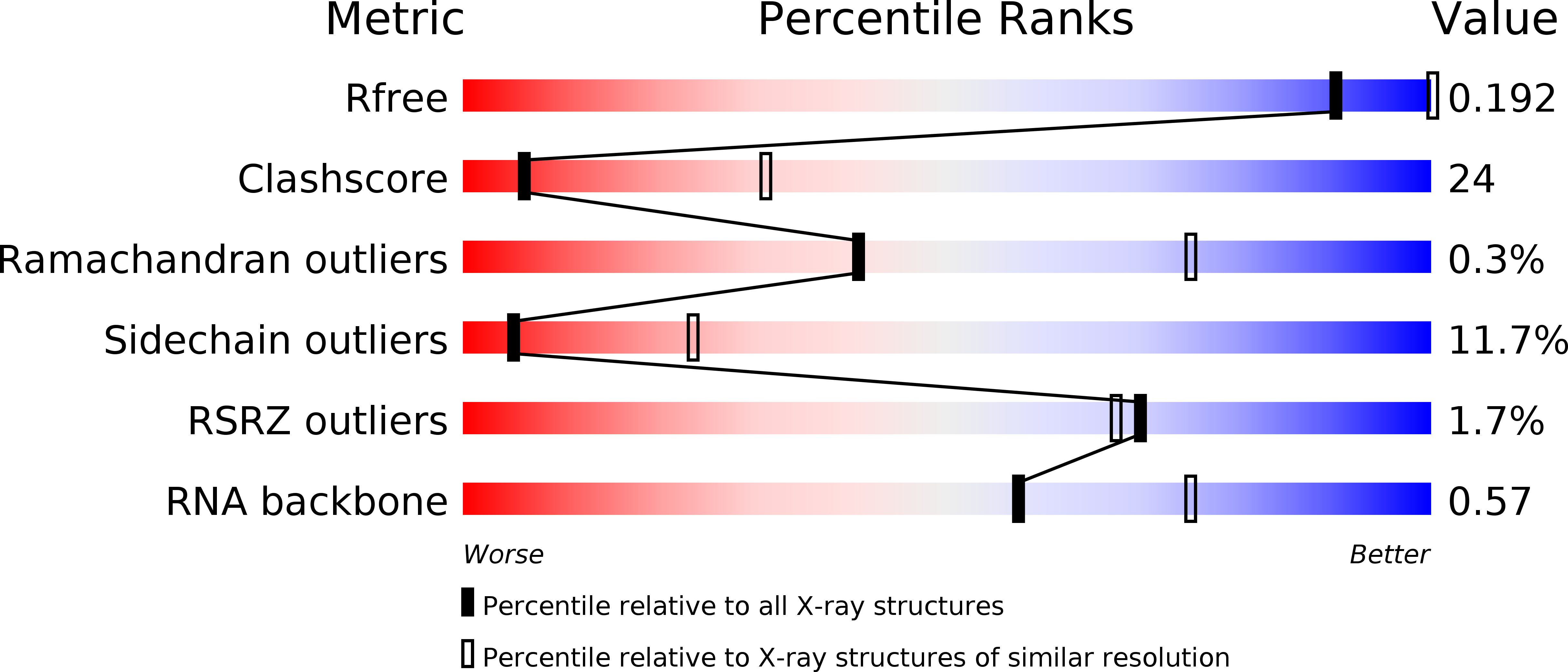
Resolution:
3.45 Å
R-Value Free:
0.19
R-Value Work:
0.15
R-Value Observed:
0.15
Space Group:
P 41 21 2
Macromolecular Entities
Polymer Type:polyribonucleotide
Description:16S rRNA
Chain IDs:A
Chain Length:1522
Number of Molecules:1
Biological Source:Thermus thermophilus
Polymer Type:polypeptide(L)
Description:30S ribosomal protein S2
Chain IDs:B
Chain Length:256
Number of Molecules:1
Biological Source:Thermus thermophilus
Polymer Type:polypeptide(L)
Description:30S ribosomal protein S3
Chain IDs:C
Chain Length:239
Number of Molecules:1
Biological Source:Thermus thermophilus
Polymer Type:polypeptide(L)
Description:30S ribosomal protein S4
Chain IDs:D
Chain Length:209
Number of Molecules:1
Biological Source:Thermus thermophilus
Polymer Type:polypeptide(L)
Description:30S ribosomal protein S5
Chain IDs:E
Chain Length:162
Number of Molecules:1
Biological Source:Thermus thermophilus
Polymer Type:polypeptide(L)
Description:30S ribosomal protein S6
Chain IDs:F
Chain Length:101
Number of Molecules:1
Biological Source:Thermus thermophilus
Polymer Type:polypeptide(L)
Description:30S ribosomal protein S7
Chain IDs:G
Chain Length:156
Number of Molecules:1
Biological Source:Thermus thermophilus
Polymer Type:polypeptide(L)
Description:30S ribosomal protein S8
Chain IDs:H
Chain Length:138
Number of Molecules:1
Biological Source:Thermus thermophilus
Polymer Type:polypeptide(L)
Description:30S ribosomal protein S9
Chain IDs:I
Chain Length:128
Number of Molecules:1
Biological Source:Thermus thermophilus
Polymer Type:polypeptide(L)
Description:30S ribosomal protein S10
Chain IDs:J
Chain Length:105
Number of Molecules:1
Biological Source:Thermus thermophilus
Polymer Type:polypeptide(L)
Description:30S ribosomal protein S11
Chain IDs:K
Chain Length:129
Number of Molecules:1
Biological Source:Thermus thermophilus
Polymer Type:polypeptide(L)
Description:30S ribosomal protein S12
Chain IDs:L
Chain Length:135
Number of Molecules:1
Biological Source:Thermus thermophilus
Polymer Type:polypeptide(L)
Description:30S ribosomal protein S13
Chain IDs:M
Chain Length:126
Number of Molecules:1
Biological Source:Thermus thermophilus
Polymer Type:polypeptide(L)
Description:30S ribosomal protein S14
Chain IDs:N
Chain Length:61
Number of Molecules:1
Biological Source:Thermus thermophilus
Polymer Type:polypeptide(L)
Description:30S ribosomal protein S15
Chain IDs:O
Chain Length:89
Number of Molecules:1
Biological Source:Thermus thermophilus
Polymer Type:polypeptide(L)
Description:30S ribosomal protein S16
Chain IDs:P
Chain Length:88
Number of Molecules:1
Biological Source:Thermus thermophilus
Polymer Type:polypeptide(L)
Description:30S ribosomal protein S17
Chain IDs:Q
Chain Length:105
Number of Molecules:1
Biological Source:Thermus thermophilus
Polymer Type:polypeptide(L)
Description:30S ribosomal protein S18
Chain IDs:R
Chain Length:88
Number of Molecules:1
Biological Source:Thermus thermophilus
Polymer Type:polypeptide(L)
Description:30S ribosomal protein S19
Chain IDs:S
Chain Length:93
Number of Molecules:1
Biological Source:Thermus thermophilus
Polymer Type:polypeptide(L)
Description:30S ribosomal protein S20
Chain IDs:T
Chain Length:106
Number of Molecules:1
Biological Source:Thermus thermophilus
Polymer Type:polypeptide(L)
Description:30S ribosomal protein THX
Chain IDs:U
Chain Length:27
Number of Molecules:1
Biological Source:Thermus thermophilus
Polymer Type:polyribonucleotide
Description:5'-R(*UP*UP*U)-3'
Chain IDs:V
Chain Length:3
Number of Molecules:1
Biological Source:Thermus thermophilus
Polymer Type:polyribonucleotide
Description:5'-R(*GP*GP*GP*AP*UP*UP*GP*AP*AP*AP*AP*UP*CP*CP*C)-3'
Chain IDs:W
Chain Length:15
Number of Molecules:1
Biological Source:Thermus thermophilus
Modified Residue
Compound ID Chain ID Parent Comp ID Details 2D Image
0TD L ASP (3S)-3-(METHYLSULFANYL)-L-ASPARTIC ACID
2MG A G 2N-METHYLGUANOSINE-5'-MONOPHOSPHATE
4OC A C 4N,O2'-METHYLCYTIDINE-5'-MONOPHOSPHATE
5MC A C 5-METHYLCYTIDINE-5'-MONOPHOSPHATE
7MG A G ?
M2G A G N2-DIMETHYLGUANOSINE-5'-MONOPHOSPHATE
MA6 A A 6N-DIMETHYLADENOSINE-5'-MONOPHOSHATE
PSU A U PSEUDOURIDINE-5'-MONOPHOSPHATE
UR3 A U 3-METHYLURIDINE-5'-MONOPHOSHATE
Primary Citation
A structural basis for streptomycin-induced misreading of the genetic code.
Nat Commun 4 1355 1355 (2013)
PMID: 23322043 DOI: 10.1038/ncomms2346

Abstact

During protein synthesis, the ribosome selects aminoacyl-transfer RNAs with anticodons matching the messenger RNA codon present in the A site of the small ribosomal subunit. The aminoglycoside antibiotic streptomycin disrupts decoding by binding close to the site of codon recognition. Here we use X-ray crystallography to define the impact of streptomycin on the decoding site of the Thermus thermophilus 30S ribosomal subunit in complexes with cognate or near-cognate anticodon stem-loop analogues and messenger RNA. Our crystal structures display a significant local distortion of 16S ribosomal RNA induced by streptomycin, including the crucial bases A1492 and A1493 that participate directly in codon recognition. Consistent with kinetic data, we observe that streptomycin stabilizes the near-cognate anticodon stem-loop analogue complex, while destabilizing the cognate anticodon stem-loop analogue complex. These data reveal how streptomycin disrupts the recognition of cognate anticodon stem-loop analogues and yet improves recognition of a near-cognate anticodon stem-loop analogue.

Legend

Protein

Chemical

Disease

Primary Citation of related structures