4DQ9 image
Deposition Date 2012-02-15
Release Date 2013-03-13
Last Version Date 2024-02-28
Entry Detail
PDB ID:
4DQ9
Title:
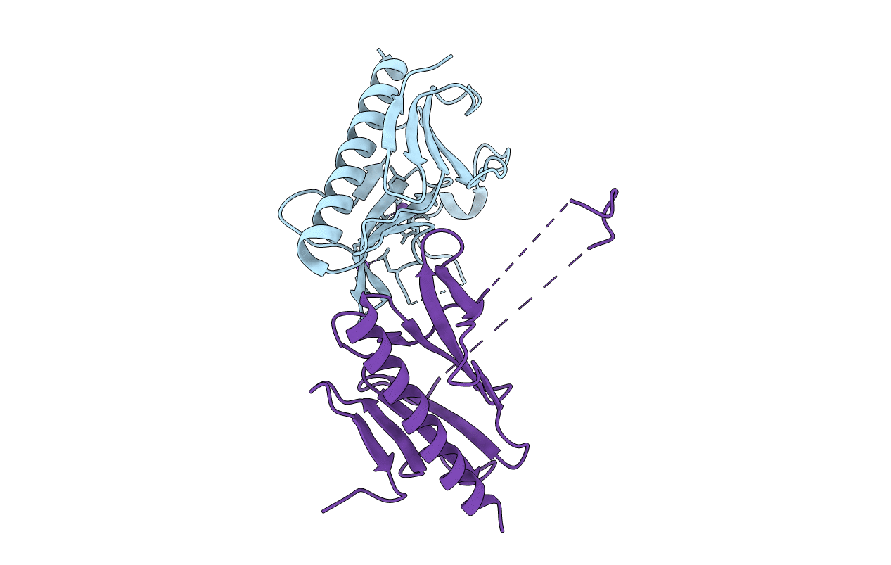
Crystal structure of the minor pseudopilin EPSH from the type II secretion system of Vibrio cholerae
Biological Source:
Source Organism:
Vibrio cholerae (Taxon ID: 666)
Host Organism:
Method Details:
Experimental Method:
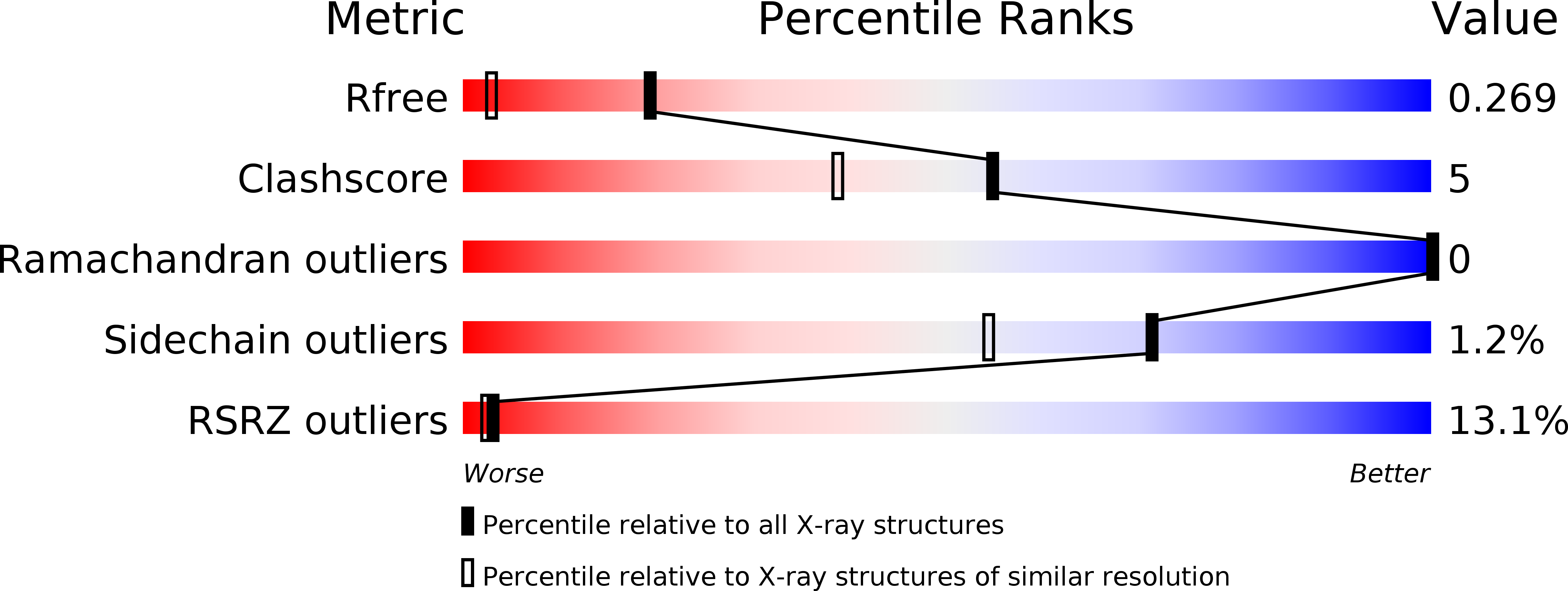
Resolution:
1.59 Å
R-Value Free:
0.24
R-Value Work:
0.20
R-Value Observed:
0.20
Space Group:
P 21 21 21
Macromolecular Entities
Structures with similar UniProt ID
Protein Blast
Polymer Type:polypeptide(L)
Molecule:General secretion pathway protein H
Gene (Uniprot):epsH
Chain IDs:A, B
Chain Length:170
Number of Molecules:2
Biological Source:Vibrio cholerae
Primary Citation
The 1.59 angstrom resolution structure of the minor pseudopilin EpsH of Vibrio cholerae reveals a long flexible loop.
Biochim.Biophys.Acta 1844 406 415 (2013)
PMID: 24316251 DOI: 10.1016/j.bbapap.2013.11.013

Abstact

The type II secretion complex exports folded proteins from the periplasm to the extracellular milieu. It is used by the pathogenic bacterium Vibrio cholerae to export several proteins, including its major virulence factor, cholera toxin. The pseudopilus is an essential component of the type II secretion system and likely acts as a piston to push the folded proteins across the outer membrane through the secretin pore. The pseudopilus is composed of the major pseudopilin, EpsG, and four minor pseudopilins, EpsH, EpsI, EpsJ and EpsK. We determined the x-ray crystal structure of the head domain of EpsH at 1.59Å resolution using molecular replacement with the previously reported EpsH structure, 2qv8, as the template. Three additional N-terminal amino acids present in our construct prevent an artifactual conformation of residues 160-166, present in one of the two monomers of the 2qv8 structure. Additional crystal contacts stabilize a long flexible loop comprised of residues 104-135 that is more disordered in the 2qv8 structure but is partially observed in our structure in very different positions for the two EpsH monomers in the asymmetric unit. In one of the conformations the loop is highly extended. Modeling suggests the highly charged loop is capable of contacting EpsG and possibly secreted protein substrates, suggesting a role in specificity of pseudopilus assembly or secretion function.

Legend

Protein

Chemical

Disease

Primary Citation of related structures