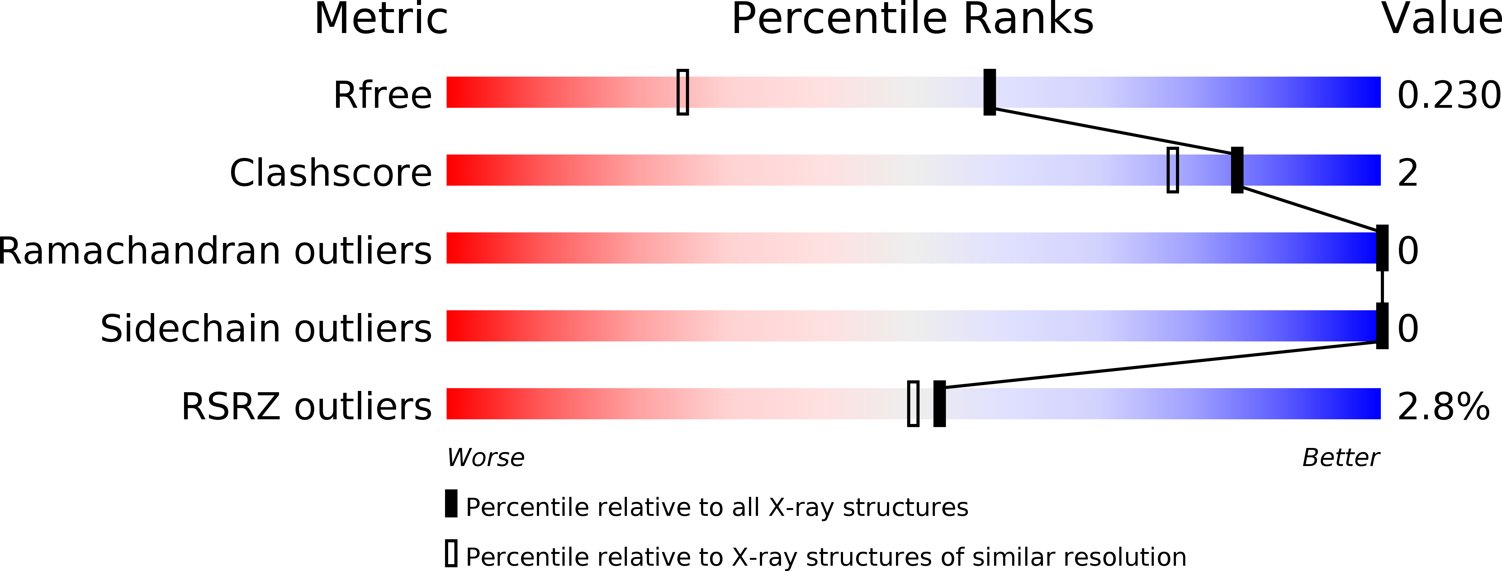
4DNY image
Deposition Date 2012-02-09
Release Date 2012-05-16
Last Version Date 2024-02-28
Entry Detail
PDB ID:
4DNY
Keywords:
Title:
Crystal structure of enterohemorrhagic E. coli StcE(132-251)
Biological Source:
Source Organism:
Host Organism:
Method Details:
Experimental Method:
Resolution:
1.61 Å
R-Value Free:
0.21
R-Value Work:
0.18
R-Value Observed:
0.18
Space Group:
H 3
Macromolecular Entities
Polymer Type:polypeptide(L)
Molecule:Metalloprotease stcE
Gene (Uniprot):stcE
Chain IDs:A
Chain Length:126
Number of Molecules:1
Biological Source:Escherichia coli
Ligand Molecules
Primary Citation
Structural Insight into the Bacterial Mucinase StcE Essential to Adhesion and Immune Evasion during Enterohemorrhagic E. coli Infection.
Structure 20 707 717 (2012)
PMID: 22483117 DOI: 10.1016/j.str.2012.02.015

Abstact

Mucin glycoproteins with large numbers of O-linked glycosylations comprise the mucosal barrier lining the mammalian gastrointestinal tract from mouth to gut. A critical biological function of mucins is to protect the underlying epithelium from infection. Enterohemorrhagic Escherichia coli (EHEC), the mediator of severe food- and water-borne disease, can breach this barrier and adhere to intestinal cells. StcE, a ∼100 kDa metalloprotease secreted by EHEC, plays a pivotal role in remodeling the mucosal lining during infection. To obtain mechanistic insight into its function, we have determined the structure of StcE. Our data reveal a dynamic, multidomain architecture featuring an unusually large substrate-binding cleft and a prominent polarized surface charge distribution highly suggestive of an electrostatic role in substrate targeting. The observation of key conserved motifs in the active site allows us to propose the structural basis for the specific recognition of α-O-glycan-containing substrates. Complementary biochemical analysis provides further insight into its distinct substrate specificity and binding stoichiometry.

Legend

Protein

Chemical

Disease

Primary Citation of related structures