Deposition Date
2012-02-08
Release Date
2012-03-28
Last Version Date
2023-09-13
Entry Detail
PDB ID:
4DMT
Keywords:
Title:
Crystal structure of a VWF binding collagen III derived triple helical peptide
Method Details:
Experimental Method:
Resolution:
1.39 Å
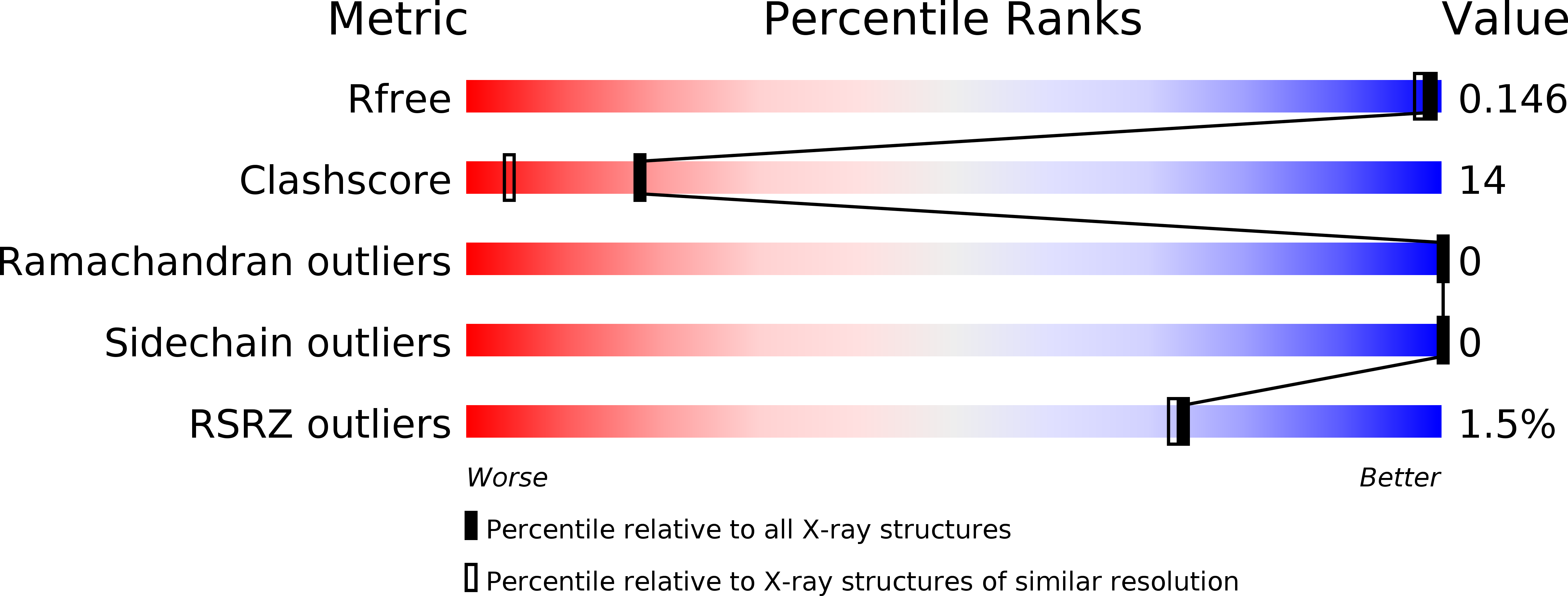
R-Value Free:
0.14
R-Value Work:
0.10
R-Value Observed:
0.10
Space Group:
P 1 21 1